Bowel-GI

To & Fro Peristalsis in Bowel Obstruction
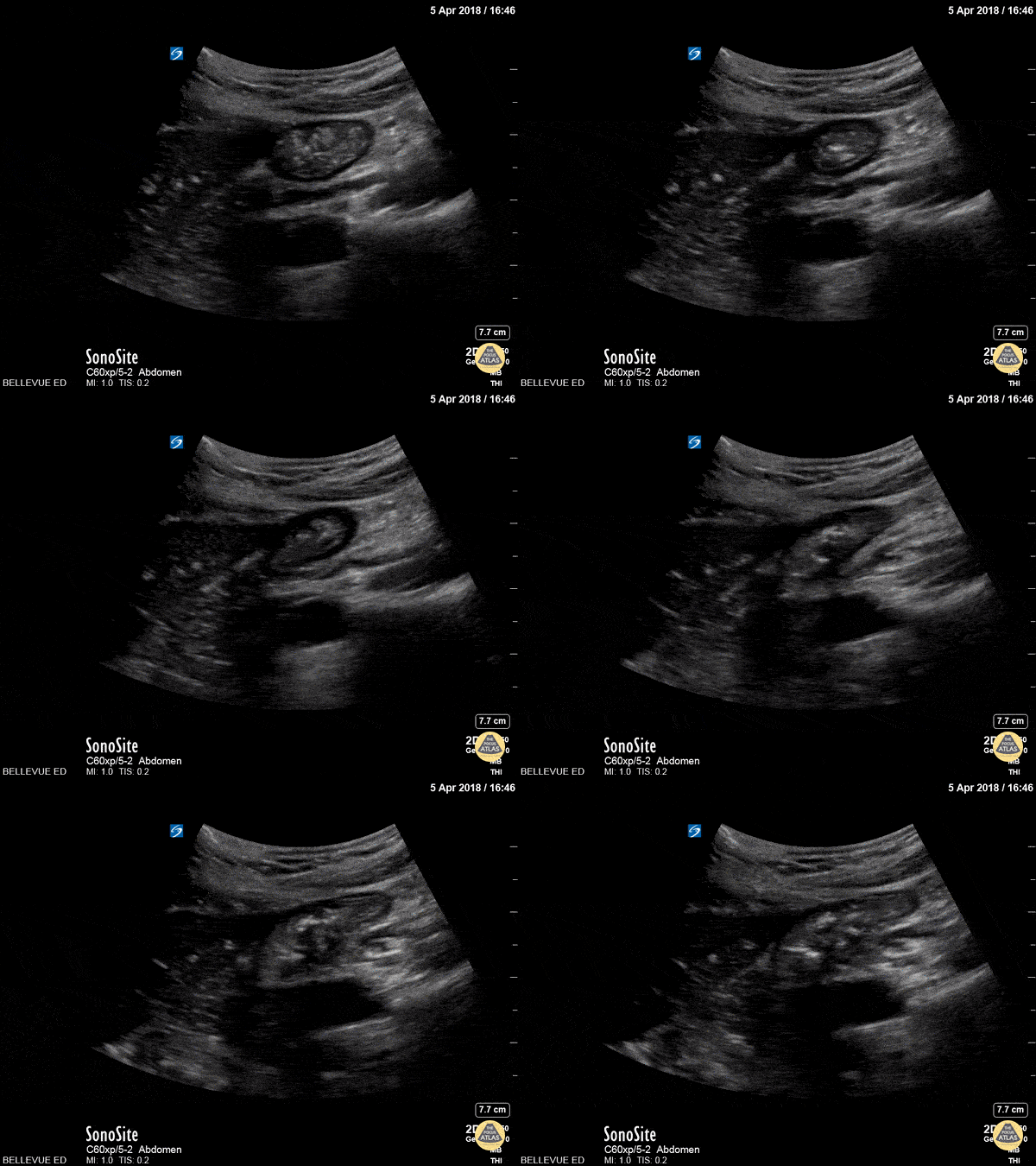
Fecal material can be seen moving forward and backwards through dilated bowels in the patient with a bowel obstruction.
Contributed by: Brittany Garza, DO and Saleem Nasseh, MD and Sadie Ellenson, MS4

SBO Demonstrating To-and-Fro Peristalsis
Middle age male patient with past history of multiple abdominal surgeries, presented with clinical picture of intestinal obstruction. POCUS was performed demonstrating dilated bowel loops up to 3.3 cm with clear "to-and-fro" movements of bowel contents on the right side of the screen.
Learning point: Keep the ultrasound probe still and wait for enough time to allow the back and forth "to-and-fro" movements of the bowel content.
Contributor: Basel Elmegabar; MBBS

Normal Bowel Peristalsis
51 year old male presented with a chief complaint of abdominal pain for 3 days with nausea and vomiting.
The curvilinear probe was used to evaluate the aorta with an incidental finding (shown) of clear peristalsis of the bowel contents with hypoechoic and anechoic contents. In this segment it is clear there is no obstruction.
Lindsay Davis, DO, MPH, @Lindsadavis18
Lydia Mansour, DO
Emily Nagourney, MS4
Central Michigan University

Small Bowel Obstruction
30-year-old presented with 2-day history of abdominal pain, abdominal distention, and vomiting. Pertinent PMH includes prior hemicolectomy due to Crohn's disease. Seen here is POCUS evidence of SBO.
Ahmad Khan
@kadiwls

SBO with "To-and-Fro"
A 58-year-old female with multiple prior abdominal surgeries presented with a 2-day hx of abdominal pain and N/V. ROS also notable for anorexia and constipation.
Abdominal POCUS seen here revealed dilated bowel loops with thickened and hyperechoic bowel walls. “To-and-fro” movements of the bowel contents are also appreciated. This occurs as a consequence of increased intestinal contents and peristalsis, otherwise known as dysfunctional peristalsis. All of the aforementioned are consistent with a diagnosis of SBO.
When there is high clinical suspicion of SBO, POCUS can serve as an inexpensive and rapid adjunct for clinical evaluation, expediting time to disposition.
Dr. Samantha Wong, @esayemDO
EM Resident. Central Michigan University

To & Fro in Bowel Obstruction
This 70-year-old male presented to our ED reporting abdominal pain and vomiting with associated absence of flatus / bowel movements. Seen here is his LUQ abdominal view using the curvilinear probe that reveals a dilated loop of bowel. The image is further characterized by increased peristalsis (the "To and Fro" sign) as well as the presence of free fluid adjacent to the dilated bowel loop in the shape of a triangle (known as the "Tanga Sign"). This case highlights how bedside ultrasound can be a powerful tool to enable prompt diagnosis and treatment of bowel obstruction.
Renato Tambelli, Emergency Physician Hospital das Clínicas de Marília, Brazil.
@R_Tambelli // @JediPocus

Duodenum
In this clip the probe is positioned over the RUQ. The liver is on the left side of the screen, and in the center of the screen we see a portion of the duodenum in cross-section. Often mistaken for a gallbladder full of gallstones by novices sonographers, it can be identified by its peristaltic waves with visible motion of the bowel contents within.
Hannah Kopinksi and Dr. Lindsay Davis - NYU Emergency Medicine

Large Bowel
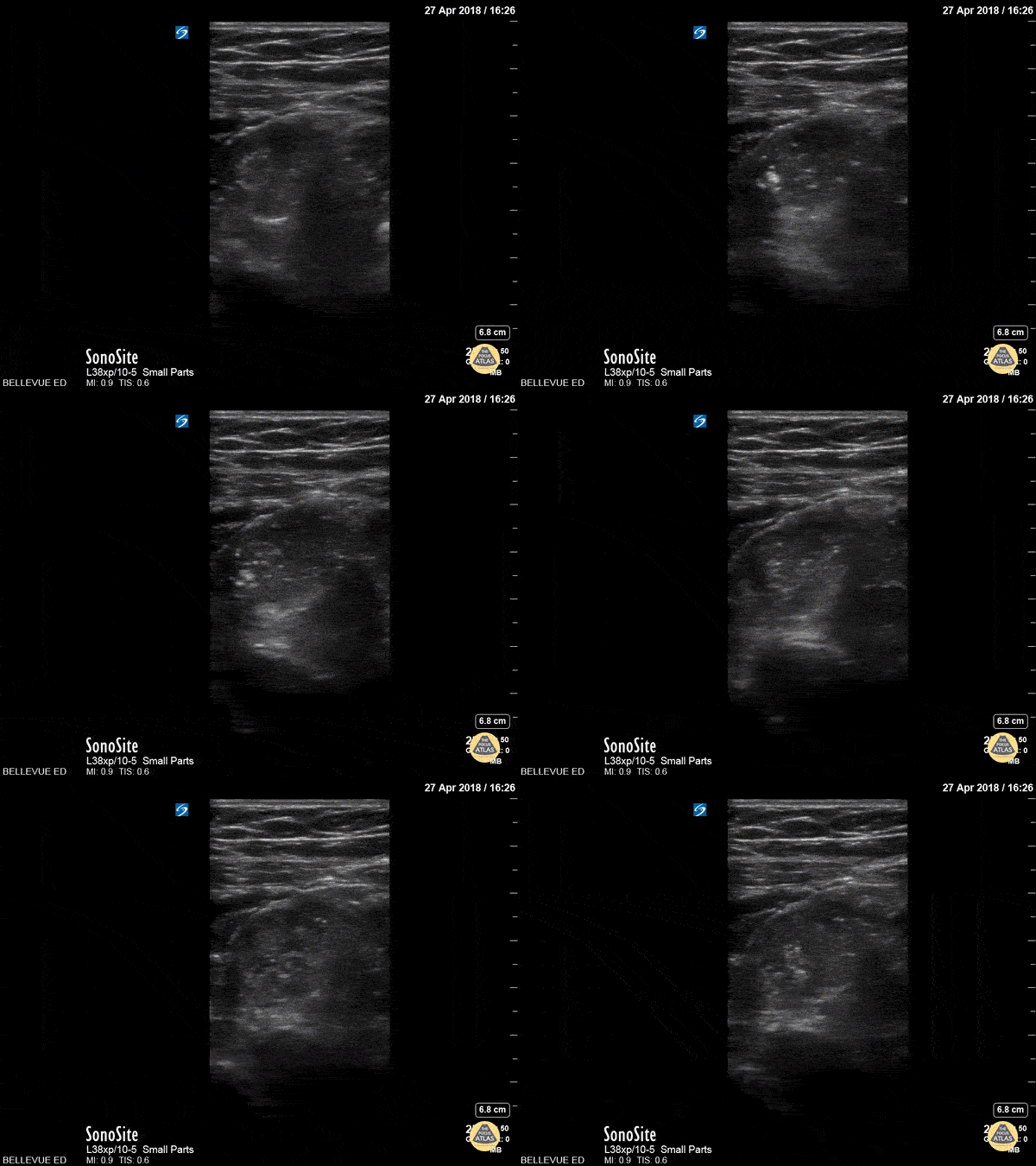
This clip shows a segment of the colon in long axis beneath layers of connective tissue and muscle. The peristalsis of the bowel is clearly visible as demonstrated by the motion of its inner contents.
Hannah Kopinksi and Dr. Lindsay Davis - NYU Emergency Medicine


