Cardiac Tumors

Cardiac Sarcoma
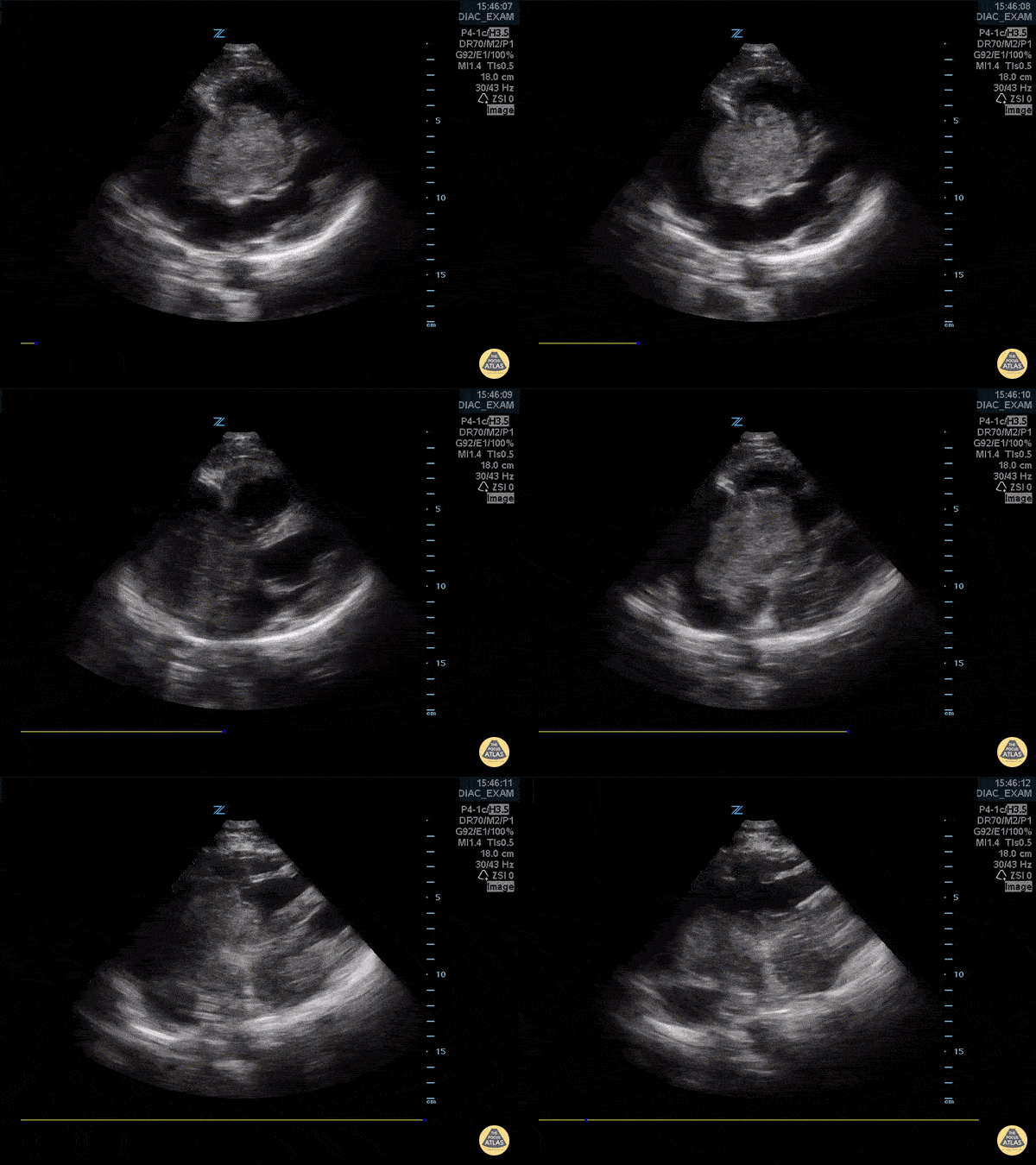
This patient presented to the emergency department after a syncopal episode upon exertion. From this subxiphoid view of their heart, we can see isoechoic structures within their right atrium. This patient was ultimately diagnosed with a cardiac sarcoma.
Image courtesy of Robert Jones DO, FACEP @RJonesSonoEM
Director, Emergency Ultrasound; MetroHealth Medical Center; Professor, Case Western Reserve Medical School, Cleveland, OH
View his original post here

Cardiac Myxoma
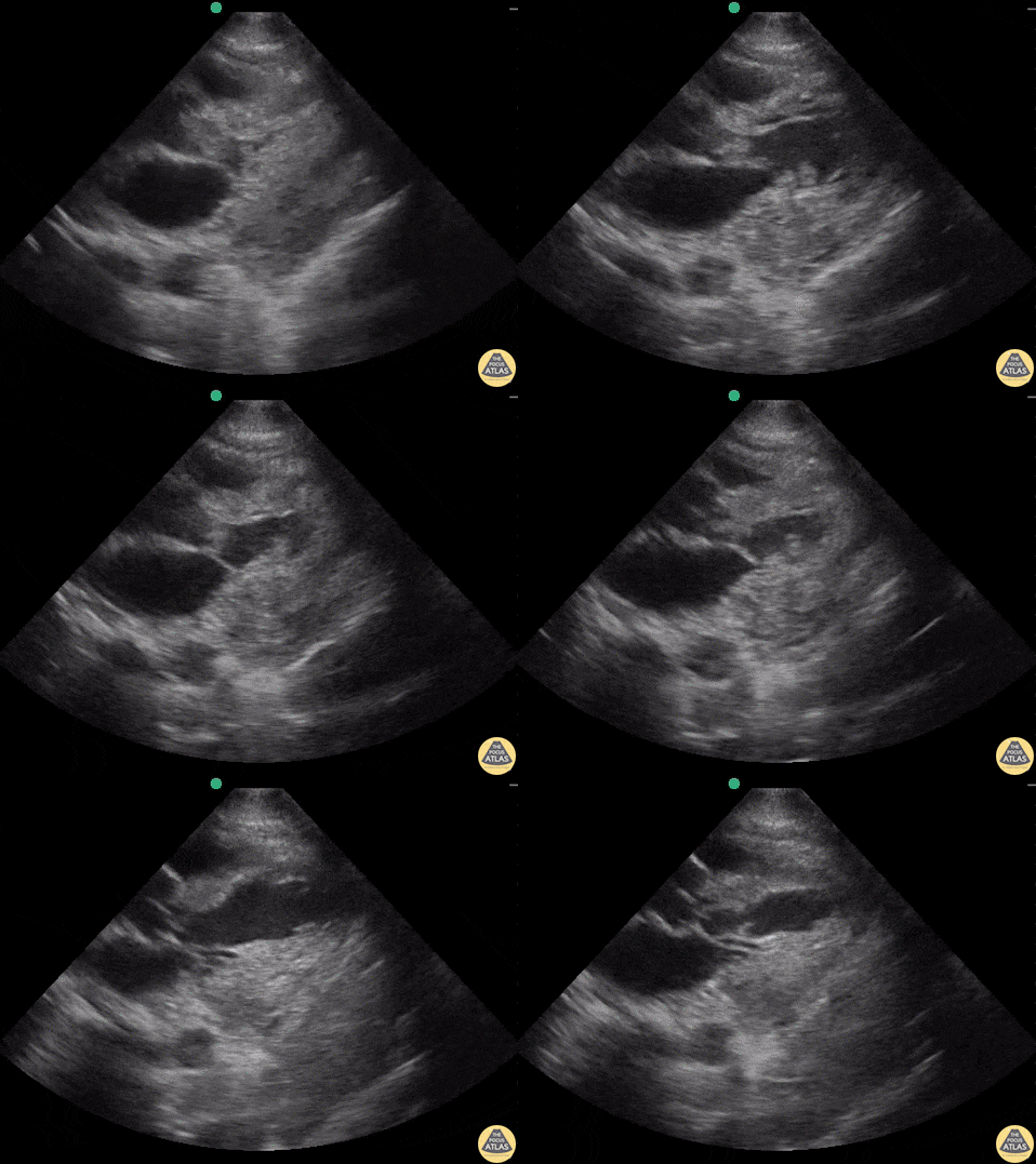
64 yo man with history of hepatic cirrhosis presented to ED with hypotension. During the RUSH exam, we incidentally identified this large hyperechoic mass occupying a large part of the right heart. Cardiac myxomas are the most common primary cardiac tumor in adults, though only 15–20% originate within the right atrium as does this one. The etiology of hypotension in our patient ended up being septic shock secondary to SBP. However, bedside ultrasound sometimes surprises you with stunning and unexpected images!
Renato Tambelli, @JediPocus
Emergency Physician (HCFAMEMA /Sao Paulo, Brazil)

Right Atrial Myxoma
An apical 4 chamber view on a patient with CP, SOB, and palpitations revealed a right sided atrial myxoma.
Image courtesy of Robert Jones DO, FACEP @RJonesSonoEM
Director, Emergency Ultrasound; MetroHealth Medical Center; Professor, Case Western Reserve Medical School, Cleveland, OH
View his original post here

Left Atrial Myxoma
Apical 4 chamber view of a patient with dyspnea of exertion revealed a left atrial myxoma.
Image courtesy of Robert Jones DO, FACEP @RJonesSonoEM
Director, Emergency Ultrasound; MetroHealth Medical Center; Professor, Case Western Reserve Medical School, Cleveland, OH
View his original post here

Atrial Myxoma
In this parasternal long axis view, a large mass is present in the right atrium that moves into the right ventricle during diastole.
Frances Russell, MD, RDMS

Cavo-Atrial Tumor-Thrombus Complex
Extensive tumor-thrombus complex originating from a right adrenal malignancy that invaded the IVC and migrated cephalad until it was prolapsing through the tricuspid valve into the RV. Shockingly, this monstrous goomba was almost invisible on PSL, PSS, and A4C views.
Submitted by Dr. Elias Jaffa

Squamous Cell Metastases to the Heart
59 y/o F PMH metastatic squamous cell carcinoma of lung with metastases to bone, brain, liver, subcutaneous tissue presents with undifferentiated SOB. Patient tachycardic, hypotensive but alert with EKG showing non-sustained ventricular tachycardia.
Multiple hypodensities and cystic lesions in the LV, proximal outflow tract in left ventricle, all suggestive of a thrombus vs mass vs vegetations. Eventual presumed diagnosis after formal transthoracic echo is metastases to the heart.
Dr. Joshua Schecter, Dr. John F. Kilpatrick - Kings County/SUNY Downstate Emergency Medicine


