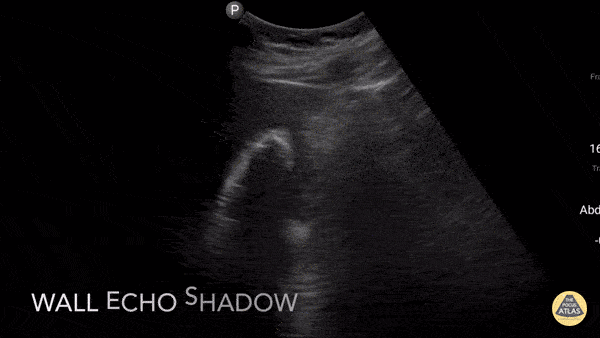
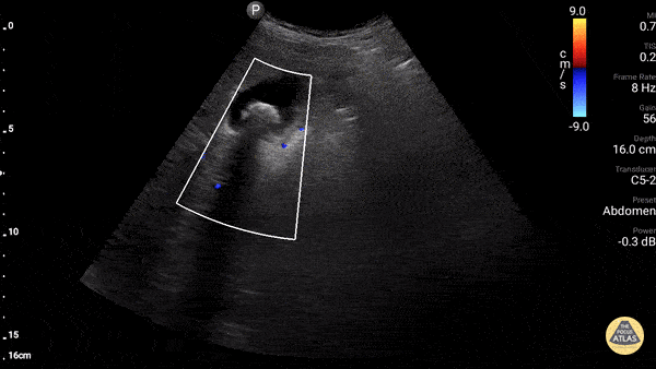
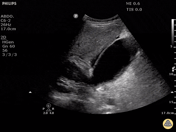
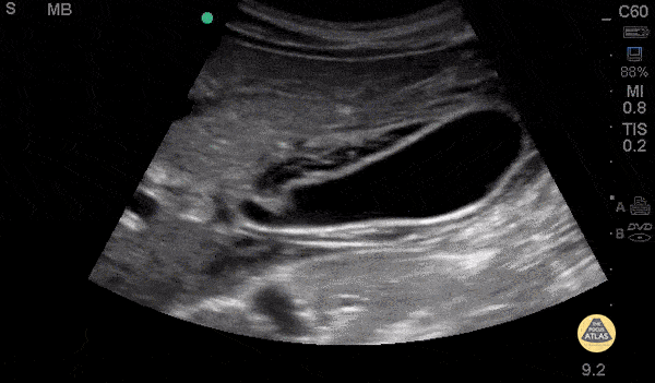
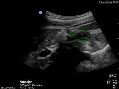
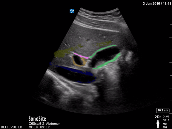
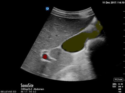
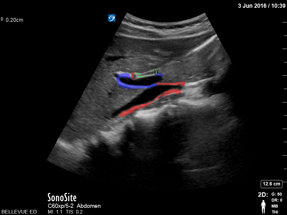
Biliary

Adenomyomatosis
Adenomyomatosis is a generally benign condition characterized by diffuse thickening of the gallbladder wall and intramural diverticula. On ultrasound it creates comet tail artifacts (reverberation artifacts that appear as tiny echogenic beams) originating from distinct locations on the gallbladder wall.
This is often seen in asymptomatic patients; however it can be associated with chronic inflammation from gallstones and pancreatitis.
Dr. Lindsay Davis - NYU/Bellevue Department of Emergency Medicine

Cholelithiasis, Not Impacted
Two large, hyperechoic stones can be appreciated in this gallbladder. These are likely two stones because of the degree of echogenicity and posterior shadowing. If you have one or two objects in the gallbladder and you're not sure if they're stones, it can be useful to have the patient lay on their side. If the objects moves when the patient moves, they are stones. If not, they are likely polyps or other masses growing from the gallbladder wall.
Justin Bowra MBBS, FACEM, CCPU Emergency Physician, RNSH et al. (Dr. Darmas)

Cholelithiasis with Many Stones
This non-inflamed gallbladder contains many stones. The stones are hyperechoic with posterior shadowing. The gallbladder wall is not thickened, there is no pericholecystic fluid, and no sonographic Murphy's sign, thus cholecystitis is unlikely.
Justin Bowra MBBS, FACEM, CCPU Emergency Physician, RNSH et al. (Dr. Ken Lee)

Cholelithiasis - Neck Stones
This patient w/o cholecystitis clearly demonstrates cholelithiasis. A 2002 study showed that ER doctors can diagnose gallstones with a sensitivity of 88% and specificity 96% with POCUS.
Gallstones can be identified by hypoechoic "shadowing" behind hyperechoic stones. If there isn't shadowing, hyperechoic structures could represent polyps or sludge. In this image the stones are resting mainly in the neck of the gallbladder.
Kendall JL, Shimp RJ. Performance and interpretation of focused right upper quadrant ultrasound by emergency physicians. J Emerg. Med. 2001; 21(1):7-13
Submitted by Justin Bowra MBBS, FACEM, CCPU Emergency Physician, RNSH et al.

Cholecystitis with Obstructing Stone
This clearly demonstrates a stone in the gallbladder neck as a hyperechoic structure with posterior shadowing. The neck is not always clearly visualized on first glance so it is important to scan through the gallbladder in two planes to exclude stones in the neck. Other signs of acute cholecystitis include pericholecystic fluid, gallbladder wall thickening (>3mm) common bile duct dilatation, and positive sonographic Murphy's sign.
Justin Bowra MBBS, FACEM, CCPU Emergency Physician, RNSH et al. (Mo)

Cholecystitis
This still image shows two clear signs of acute cholecystitis. The gallbladder wall is thickened and there is pericholecystic fluid. Of note, when measuring the gallbladder wall it's better to measure the anterior wall as the posterior wall can appear enlarged due to artifact. The cut off for wall thickening is 3mm. The anterior wall is thickened in this case but is not labeled. Other signs to look for are stones, sludge, common bile duct dilatation, and overall gallbladder enlargement.
Of note, a positive sonographic Murphy's carries a sensitivity of 63%, specificity of 93.6% and positive predictive value of 72.5%.
Justin Bowra MBBS, FACEM, CCPU Emergency Physician, RNSH et al. (Sharon)

Cholecystitis - Emphysematous
Emphysematous cholecystitis is the result of gas formation within the wall or lumen of the gallbladder as a result of infection by a gas forming organism. It is a surgical emergency. The air in the gallbladder appears highly echogenic with mild posterior shadowing, as air next to tissue is highly reflective.
Sukh Singh, MD

Choledocholithiasis - Dilated Common Bile Duct with Stone
A normal common bile duct should be < 4mm plus 1mm per decade after 40 years of age. A stone can clearly be seen in the area of color flow doppler. The bile duct is obviously dilated here (although not measured).
When searching for the CBD, it can be useful to turn color flow doppler on to ensure you're visualizing a biliary structure and not vessels. This technique was employed here.
POCUS is highly sensitive for acute cholecystitis but as many people already know, it can be hard to find the CBD, which is reflected in the lower sensitivity and specificity for this indication in many studies.
Justin Bowra MBBS, FACEM, CCPU Emergency Physician, RNSH et al. (Dr. Dop Ahilan)

Common Bile Duct (Normal)
When searching for the common bile duct (CBD), color doppler can be useful to differentiate vascular structures from non-vascular structures. In this case the CBD is being measured anteriorly and does not have any pulsations on color doppler. The portal vein and artery can be seen and with the CBD, make the portal triad.
A normal CBD should be less than 4mm plus 1mm per decade after 40 years old. It should be measured inner wall to inner wall.
Justin Bowra MBBS, FACEM, CCPU Emergency Physician, RNSH et al. (Dr. Chiang)

Common Bile Duct (Normal Labeled)
A normal common bile duct should be <4mm plus 1mm per decade after 40 years of age. It should be measured from inner wall to inner wall.
You should attempt to find the portal triad: the CBD, the portal vein, and the hepatic artery. You may need color flow to find all three. Here the CBD and portal vein are labeled (no labeled hepatic artery).
To get better images, especially if you're dealing with a lot of rib shadow, it can be helpful to have your patient take a deep breath and/or roll onto their left side.
Justin Bowra MBBS, FACEM, CCPU Emergency Physician, RNSH et al.

Dilated Common Bile Duct
The CBD measures at 1.89cm indicating it is grossly dilated, as a normal common bile duct should be <4mm plus 1mm per decade after 40 years of age. It should be measured from inner wall to inner wall. The color doppler is used to ensure that the dilated structure is indeed a bile duct rather than a vascular abnormality.
Sukh Singh, MD

Dilated Common Bile Duct
This patient has a dilated common bile duct and obstructive pattern LFTs, highly suggestive of choledocholithiasis or other biliary obstruction. No stone is visualized on this study.
A normal common bile duct should be <4mm plus 1mm per decade after 40 years of age. Here it is 9.78 mm.
POCUS is highly sensitive for acute cholecystitis but as many people already know, it can be hard to find the CBD, which is reflected in the lower sensitivity and specificity for this indication in many studies.
Justin Bowra MBBS, FACEM, CCPU Emergency Physician, RNSH et al. (Dr. Ken Lee)

Gallbladder Wall Measurement
This normal gallbladder image shows how to measure the anterior wall of the gallbladder. A normal gallbladder wall should be less than 3mm. The posterior wall should not be measured as it can appear enlarged due to artifact.
Justin Bowra MBBS, FACEM, CCPU Emergency Physician, RNSH et al.

Gallbladder with Biliary Sludge
The image above is from a patient presenting to the emergency department with recurrent abdominal pain. The image demonstrates a large amount of biliary sludge within a normal sized gallbladder. Biliary colic from intermittent obstruction was suspected. While the gallbladder wall is noted to be mildly thick here, cholecystitis was ruled out based on other clinical factors.
Marco Garrone, MD - Torino, Italy

Gallbladder Sludge
Echogenic material can be seen within this gallbladder but there are no stones. It is not bright white (it is not highly echogenic) and furthermore there isn't any shadowing. When there is no shadowing consider polyps or in this case biliary sludge.
Justin Bowra MBBS, FACEM, CCPU Emergency Physician, RNSH et al. (Sharon)

Gallbladder Wall Echo Shadow (WES Sign)
Gallstones can be seen on the right side of the image with a hyperechoic front edge and posterior shadowing.
Justin Bowra MBBS, FACEM, CCPU Emergency Physician, RNSH et al.

Wall Echo Shadow
Pt with epigastric pain, elevated LFTs and lipase. U/S shows the whole gallbladder shadowed by a large calcified stone or "wall echo shadow-WES." The novice may not be able to find the GB and can mistake WES for bowel (but bowel does not shadow like this).
Dr. Gordon Johnson

Portal Triad
Gallbladder with non-obstructing stone. Fanning down one sees the portal triad with bile duct (black) 7 mm, hepatic artery (pulsitile) and portal vein (red).
Dr. Gordon Johnson

Pericholecystic Fluid
Free fluid can be appreciated around this gallbladder. This is called pericholecystic fluid. However there are no stones, nor gallbladder wall thickening, and the sonographic Murphy's is negative. This free fluid is unlikely to be caused by cholecystitis. In this case, the free fluid came from a different source.
Justin Bowra MBBS, FACEM, CCPU Emergency Physician, RNSH et al.

Pericholecystic Fluid - Long
26 y/o F PMH HIV presents with non-bloody, non-bilious vomiting for one day associated with upper abdominal pain. POCUS revealed gallbladder wall thickening and pericholecystic fluid, but no gallstones or sonographic Murphy’s sign. The patient received symptomatic treatment and a surgical evaluation. The patient ultimately improved and was determined to not have cholecystitis. The patient was discharged home after her symptoms resolved and she was able to tolerate PO.
Dr. Guru Shan, Dr. Guy Carmelli, Dr. Scott Kendal - Kings County Emergency Medicine

Pericholecystic Fluid - Transverse
26 y/o F PMH HIV presents with non-bloody, non-bilious vomiting for one day associated with upper abdominal pain. POCUS revealed gallbladder wall thickening and pericholecystic fluid, but no gallstones or sonographic Murphy’s sign. The patient received symptomatic treatment and a surgical evaluation. The patient ultimately improved and was determined to not have cholecystitis. The patient was discharged home after her symptoms resolved and she was able to tolerate PO.
Dr. Guru Shan, Dr. Guy Carmelli, Dr. Scott Kendal - Kings County Emergency Medicine

Free Fluid around Normal Gallbladder
There is a lot of pericholecystic fluid surrounding this gallbladder but no other signs of inflammation. The gallbladder wall is normal, there are no stones, and there is no sonographic Murphy's sign. The free fluid is likely from another source, not acute cholecystitis.
Justin Bowra MBBS, FACEM, CCPU Emergency Physician, RNSH et al.

Pancreatic Pseudocyst
40s F PMH severe ETOH use disorder and ETOH pancreatitis as well as recent COVID diagnosis presented with continued dyspnea and sore throat as well as abdominal bloating with a 15-20lb weight loss in 2 months. POCUS of the upper abdomen is shown here, demonstrating a large pseudocyst, which was confirmed on CT. The patient was admitted for her symptoms and was seen by GI but ultimately was planned for outpatient drainage rather than urgent/emergent.
Dr. Nimish Bhatt, Ultrasound Fellow
Denver Health Ultrasound Fellowship

Impacted Gallbladder Neck Stone
20s F presented with recurrent postprandial RUQ pain. POCUS demonstrated multiple hyperechoic stones in the gallbladder including one in the gallbladder neck. No pericholecystic fluid or wall thickening were noted, and the common bile duct was normal caliber. Formal RUQ US confirmed these findings, and the patient was taken to the OR by the surgery team for cholecystectomy.
Dr. Nhu-Nguyen Le, Ultrasound Fellow
Denver Health Ultrasound Fellowship

Contracted Gallbladder - Colorized
Contracted Gallbladder
Green- Gallbladder
Images: Dr. Lindsay Davis, Dr. Hannah Kopinski. Image Editing: Michael Amador and Dr. Matthew Riscinti

Gallbladder and Commmon Bile Duct - Colorized
Gallbladder and Common Bile Duct
Green: Gallbladder, Blue: IVC, Orange: Portal vein, Pink: Common bile duct, Yellow: Lumen of hepatic vein
Images: Dr. Lindsay Davis, Dr. Hannah Kopinski. Image Editing: Michael Amador and Dr. Matthew Riscinti

Normal Gallbladder - Colorized
Normal Gallbladder
Yellow: Lumen of gallbladder, Red: Portal vein, Green: Common bile duct
Images: Dr. Lindsay Davis, Dr. Hannah Kopinski. Image Editing: Michael Amador and Dr. Matthew Riscinti

Olive Sandwich Sign - Colorized
Olive Sandwich Sign
Red (small): “Olive” or hepatic artery, Green: Common bile duct, Blue: Portal vein, Red (large): IVC
Images: Dr. Lindsay Davis, Dr. Hannah Kopinski. Image Editing: Michael Amador and Dr. Matthew Riscinti


