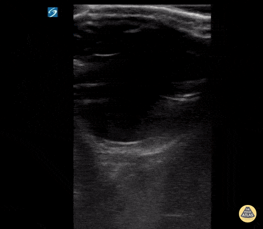
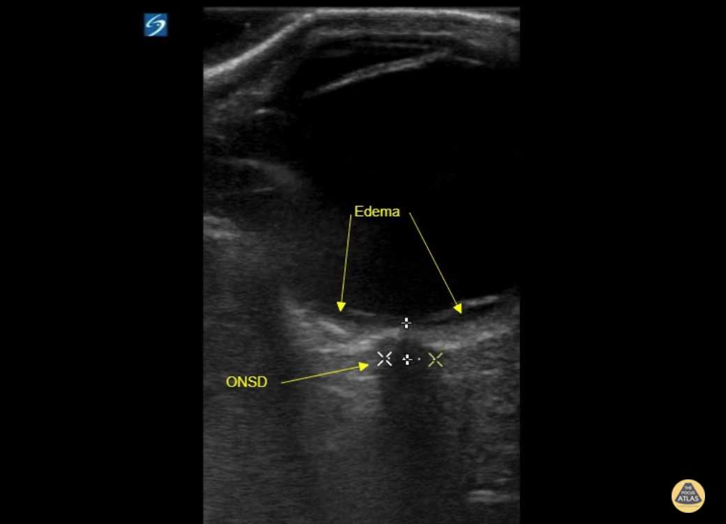
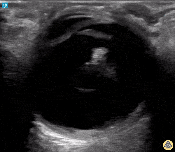
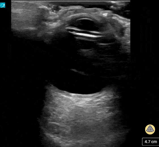
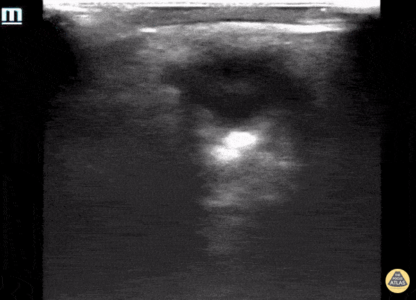
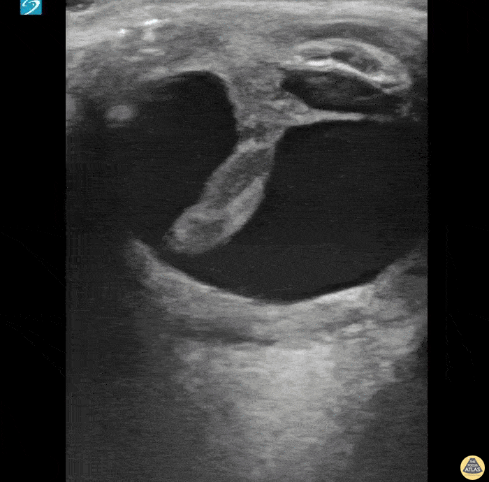
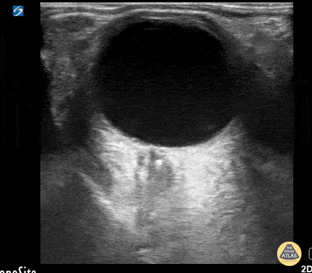
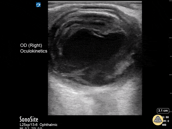
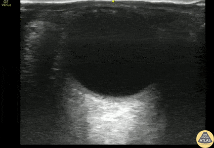
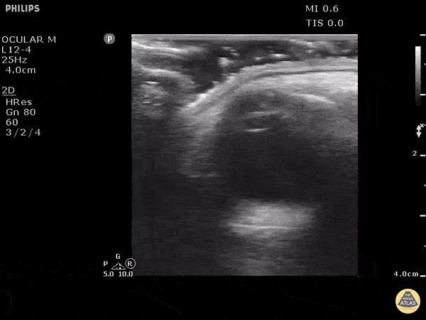
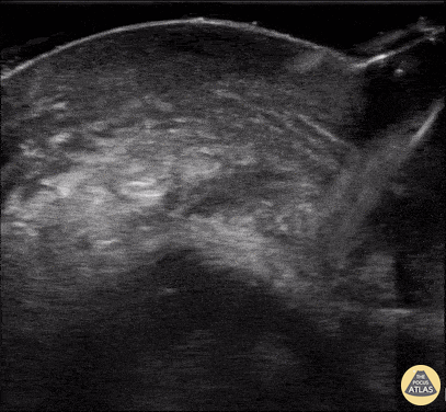
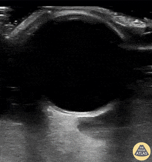
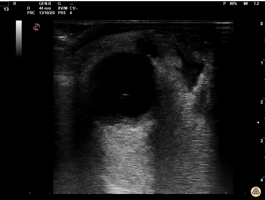
Orbital

Retinal Edema (2/2 - Clip)
17 y/o female with sudden onset bilateral blurry vision after prolonged indoor cat exposure. Fundoscopy showed retinal pigment epithelium loss consistent with bilateral neuroretinitis from cat scratch disease (Bartonella henselae).
The hyperechoic retina, normally flush with the posterior aspect of the globe, is seen separated by a hypoechoic band of edema.
Retinal edema from cat scratch disease is rare (Gen Opth 19th ed. 2017:378-422), but can be diagnosed by POCUS.
Contributed by: John Hipskind, MD, Ultrasound Director, Kaweah Health EM Residency

Retinal Edema (1/2 - Still)
17 y/o female with sudden onset bilateral blurry vision after prolonged indoor cat exposure. Fundoscopy showed retinal pigment epithelium loss consistent with bilateral neuroretinitis from cat scratch disease (Bartonella henselae).
The hyperechoic retina, normally flush with the posterior aspect of the globe, is seen separated by a hypoechoic band of edema.
Retinal edema from cat scratch disease is rare (Gen Opth 19th ed. 2017:378-422), but can be diagnosed by POCUS.
Contributed by: John Hipskind, MD, Ultrasound Director, Kaweah Health EM Residency

Intraoccular Lens Subluxation
A 50 year old male with a prior history of bilateral intraoccular lens (IOL) transplants presented to our ED with sudden onset foggy vision in his right eye while getting out of the shower. He was unable to participate in visual acuity due to the extent of his blurred. POCUS demonstrated “iridodonesis” and a provisional diagnosis of IOL subluxation/dislocation was made. Ophthalmology was consulted and confirmed the diagnosis.
Dr. Piaseczny, PGY4 Emergency Medicine, Queen's University, Kingston, Ontario, Canada

Prosthetic Ocular Lens Subluxation
This patient presented with decreased vision on a background of advanced macular degeneration. VA in the eye had decreased from 20/150 to 20/400 on presentation.
Interestingly the patient stated "I think my ocular lens has displaced."
Contributed by: Colin Bell, FRCPC, DPSPC

Phthisis Bulbi secondary to end stage eye disease
93 year old male with established end stage eye disease found incidentally to have prominent Phthisis Bulbi made evident by a heterogeneous hyper-echoic structures (calcifications) within the posterior chamber of the left eye.
Contributed by: Davis, Lindsay; Schlangen, Alex; Welch, Matthew

Old Globe Rupture
Irregularly shaped eye with internal dense structure concerning for intraocular hematoma. This patient had a history of previous rupture of the globe.
A CT Head without IV contrast described the left eye as follows: Small left globe with high density concerning for intraocular hemorrhage. Direct examination is recommended.
Halimah Hamidu-Egiebor Central Michigan University College of Medicine MD-MBA '24, Arthur Sieron Central Michigan University Emergency Medicine Resident, Eric 'Dax' Spencer Central Michigan University Emergency Medicine Resident

Central Retinal Artery Occlusion
This is from a 63 year old female who initially presented to the emergency department with vision loss over the last 24 hours. She reported bending over when she experienced complete vision loss from one of her eyes. Point of care ultrasound was performed, locating the optic nerve, but more interestingly a hyperechoic structure within the nerve (spot sign).
Overall, this is was suggestive of a central retinal artery occlusion. In this sort of situation, color doppler can also be utilized to assess for arterial versus venous occlusion.
Dr. Christopher Paulo, DO, PGY-1
Riverside Regional Medical Center Emergency Medicine Program (Newport News, VA)

Orbital Endophthalmitis
A 43 year old male with 3 days of painful right eye vision loss, fever, and URI symptoms presented to the emergency department. POCUS demonstrates echogenic swirl within the vitreous cavity. The patient was diagnosed with endogenous endophthalmitis caused by Klebsiella Pneumoniae.
Contributor: Hyun J. Yi (John), D.Sc, PA-C, MAJ, USA, Emergency Ultrasound Fellow, Madigan Army Medical Center, Joint Base Lewis-McChord, WA

Lens Subluxation
Middle aged male with prior bilateral lens replacement presented with atraumatic vision loss. POCUS performed that demonstrates subluxed lens without retinal detachment. Discussed with ophthalmology and pt was discharged with 24 hour follow up with ophthalmology, with eventual plan for operative repair.
Michael Maurantonio, PGY3, Denver Health Residency in Emergency Medicine
Michael Del Valle, Fellow, Denver Health Ultrasound Fellowship

Metallic Foreign Body In Orbital Soft Tissue
This is a clip from a patient presenting with foreign body sensation. They had a piece of metal hit their eye a week ago. Previously seen for complaint but told no foreign body seen on exam.
On ultrasound clip a hyperechoic structure can be seen adjacent to the globe. Note that with dynamic eye movements the foreign body does not move suggesting this is outside of the globe.
Image courtesy of Robert Jones DO, FACEP @RJonesSonoEM
Director, Emergency Ultrasound; MetroHealth Medical Center; Professor, Case Western Reserve Medical School, Cleveland, OH
View his original post here

Ruptured Globe
This patient sustained blunt trauma to the eye from a fireworks injury. CT scan performed prior to POCUS showed intra-orbital hematoma and complete absence of any globe tissue. The patient was taken to the OR for primary enucleation, washout, and closure.
Earl “Quinn” Cummings, MD @resus_bae
Assistant Professor; MUSC Emergency Medicine/Ultrasound; MUSC Pediatric Emergency Medicine

Retrobulbar Hematoma
Patient experienced ocular trauma 3 days prior to this exam, having been struck with a baseball to the eye. Notice the hypoechoic area within the retrobulbar space, consistent with a retrobulbar hematoma.
Tomasz Przednowek & Jereme Long @jplongest

Endogenous Endophthalmitis
A middle aged female with ESRD and longstanding percutaneous HD catheter with recent MSSA bacteremia was admitted with septic shock. She subsequently developed subacute bilateral visual loss (OS > OD). Clinical suspicion of endogenous endophthalmitis was initially supported by POCUS notable for heterogenous intraoccular material within vitreous. She was immediately started in intravitreal antibiotics in addition to previously initiated systemic antibiotics. Diagnosis of endogenous endophthalmitis was subsequently confirmed by vitrectomy.
Tessa W. Damm, DO
Intensivist, Critical Care Medicine & Neurocritical Care.
Wisconsin, USA.
@DrDamm

Asteroid hyalosis (an ultrasonographic mimic of vitreous hemorrhage)
An 59-year-old male with PMH of ESRD presented with 10-day hx floaters in his right eye. Sonographic findings are shown, notable for multiple, mobile, hyperechoic densities that swirled rapidly with eye movement. The appearance is similar to that of vitreous hemorrhage.
The patient was subsequently evaluated by ophthalmology who confirmed a diagnosis of asteroid hyalosis; a rare, benign condition of calcium phospholipid deposition within the vitreous fluid. The sonographic findings are so similar to vitreous hemorrhage that the two are commonly mistaken for each other. The clinical differentiation is that asteroid hyalosis is most often asymptomatic, and almost always without visual deficits (possibly benign floaters). These patients also rarely require vitrectomy.
In addition to clinical presentation, asteroid hyalosis can be differentiated from vitreous hemorrhage by subtle sonographic features. In asteroid hyalosis, the hyperechoic calcium phospholipid particles have a sparkling, "starry sky" appearance compared to the typically duller heterogenous blood seen in vitreous hemorrhage.
Vicky Lam, MD, MS; Christianna Sim, MD; Olusola Sanusi, MD
Kings County Hospital, SUNY Downstate Emergency Medicine

Retrobulbar Hematoma
A 65-year-old patient presented with sudden-onset headache with associated right eye pain and diplopia. Physical exam also notable for an unsteady gait, exophthalmus, and impaired EOM including absent upward and medial gaze of right eye. Orbital POCUS revealed a retrobulbar hematoma causing external compression of the optic nerve.
Marco Garrone, Emergency Medicine Physician
@drmarcogarrone

Lense Replacement
60 year-old woman with longstanding history of severe cataracts presenting 5-months s/p cataract extraction with intraocular lens placement (CEIOL). Ultrasound exam demonstrates a linear echogenic structure posterior to the iris with reverberation artifact in place of normal lenticular structure (native lens) consistent with artificial lens.
Image acquired by Robert J. McMickle (Medical Student IV, UCLA) under guidance of Yiju Teresa Liu, M.D., RDMS at Harbor-UCLA Medical Center.

Orbital Abscess
Orbital cellulitis evolving into abscess in diabetic patient. Multiple pus collections are visible on the medial side.
Dr. Marco Garrone

Lens Dislocation
Patient with severe maxillofacial trauma of unclear etiology. Ocular POCUS demonstrates a biconvex structure with a hyperechoic rim consistent with traumatic lens dislocation.
Dr. Joshua Schechter - SUNY Downstate/Kings County Emergency Medicine

Central Retinal Artery Occlusion
Only venous flow can be appreciated in this color flow image. The central retinal artery has been occluded.
Dr. Joshua Schechter - SUNY Downstate/Kings County Emergency Medicine

Subcutaneous Emphysema - Eyelid
Eyelid subcutaneous emphysema detected on US ocular scan
Dr. Marco Garrone


