Orbital

Vitreous Detachment
This was from a 52 year old male with a recent surgical history of vitreous detachment that became a retinal detachment. He presented to the emergency room for vision changes in same eye after an object fell and hit his head. Ocular ultrasound was performed at the bedside which revealed a new vitreous detachment. Important to note here that it is crucial to fan/tilt through the entire eye, making sure to visualize the optic nerve in order to differentiate between a vitreous and retinal detachment. In this case, notice how the lesion spotted in the chamber does not connect to the optic nerve, which is consistent with a vitreous detachment.
Dr. Christopher Paulo, DO, PGY-1
Riverside Regional Medical Center Emergency Medicine Program (Newport News, VA)

Vitreous Hemorrhage
An 81 yo female on ASA presented with 6 hour hx of blurred vision in her right eye. Ocular POCUS demonstrated a hyperechoic clot, seen here as 'swirling' movement, with debris settling near the bottom of the globe when eye is not in motion (end of clip). In early presentation of vitreous hemorrhage when blood is still liquified, increasing the ultrasound gain can greatly assist with identification of pathology.
Mandy Peach, MD @mandy_peach
Saint John Regional Hospital. NB, Canada

Vitreous Hemorrhage due to Proliferative Diabetic Retinopathy
A 70-year-old man presented with loss of vision to his right eye (hand motion and light perception). He takes warfarin and has diabetes. Monocular vision loss triggered a POCUS that revealed a hazy echogenic substance (consistent with blood) seen best when patient activates extra-ocular muscle movements. Also note the associated bright linear structure within one portion of the bleed, the hyaloid membrane. This patient was subsequently assessed by the Eye Hospital and diagnosed with vitreous hemorrhage due to proliferative diabetic retinopathy.
Peter Cheng

Vitreous Hemorrhage
A 71-year-old male with a history of hypertension and diabetes presented to the ED reporting gradual vision loss in his right eye over a 3-day period. Physical exam was notable for a 3 mm and nonreactive pupil with ipsilateral visual acuity reduced to patient only being able to detect movement. POCUS revealed swirling echodensity within the right eye, most appreciable with extraocular muscle movements. Ophthalmology subsequently confirmed our suspected diagnosis of vitreous hemorrhage.
Richard Cunningham, MD @HappyDays_EM
Maricopa Medical Center

Endogenous Endophthalmitis
A middle aged female with ESRD and longstanding percutaneous HD catheter with recent MSSA bacteremia was admitted with septic shock. She subsequently developed subacute bilateral visual loss (OS > OD). Clinical suspicion of endogenous endophthalmitis was initially supported by POCUS notable for heterogenous intraoccular material within vitreous. She was immediately started in intravitreal antibiotics in addition to previously initiated systemic antibiotics. Diagnosis of endogenous endophthalmitis was subsequently confirmed by vitrectomy.
Tessa W. Damm, DO
Intensivist, Critical Care Medicine & Neurocritical Care.
Wisconsin, USA.
@DrDamm

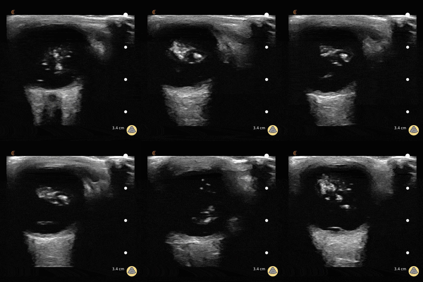
Asteroid hyalosis (an ultrasonographic mimic of vitreous hemorrhage)
An 59-year-old male with PMH of ESRD presented with 10-day hx floaters in his right eye. Sonographic findings are shown, notable for multiple, mobile, hyperechoic densities that swirled rapidly with eye movement. The appearance is similar to that of vitreous hemorrhage.
The patient was subsequently evaluated by ophthalmology who confirmed a diagnosis of asteroid hyalosis; a rare, benign condition of calcium phospholipid deposition within the vitreous fluid. The sonographic findings are so similar to vitreous hemorrhage that the two are commonly mistaken for each other. The clinical differentiation is that asteroid hyalosis is most often asymptomatic, and almost always without visual deficits (possibly benign floaters). These patients also rarely require vitrectomy.
In addition to clinical presentation, asteroid hyalosis can be differentiated from vitreous hemorrhage by subtle sonographic features. In asteroid hyalosis, the hyperechoic calcium phospholipid particles have a sparkling, "starry sky" appearance compared to the typically duller heterogenous blood seen in vitreous hemorrhage.
Vicky Lam, MD, MS; Christianna Sim, MD; Olusola Sanusi, MD
Kings County Hospital, SUNY Downstate Emergency Medicine

Vitreous Hemorrhage - Washing Machine Sign
47 y/o M with coronary artery disease on aspirin/clopidogrel with 4 days of decreased vision in his right eye. POCUS demonstrates swirling, amorphous echogenicities known as "the washing machine sign." If the eye remains still the blood will settle with gravity.
Fundoscopic evaluation by ophthalmology confirmed the diagnosis of vitreous hemorrhage.
Dr. Matthew Riscinti and Dr. Taylor Conrad - Kings County/SUNY Downstate Emergency Medicine


