Orbital

Vitreous Detachment
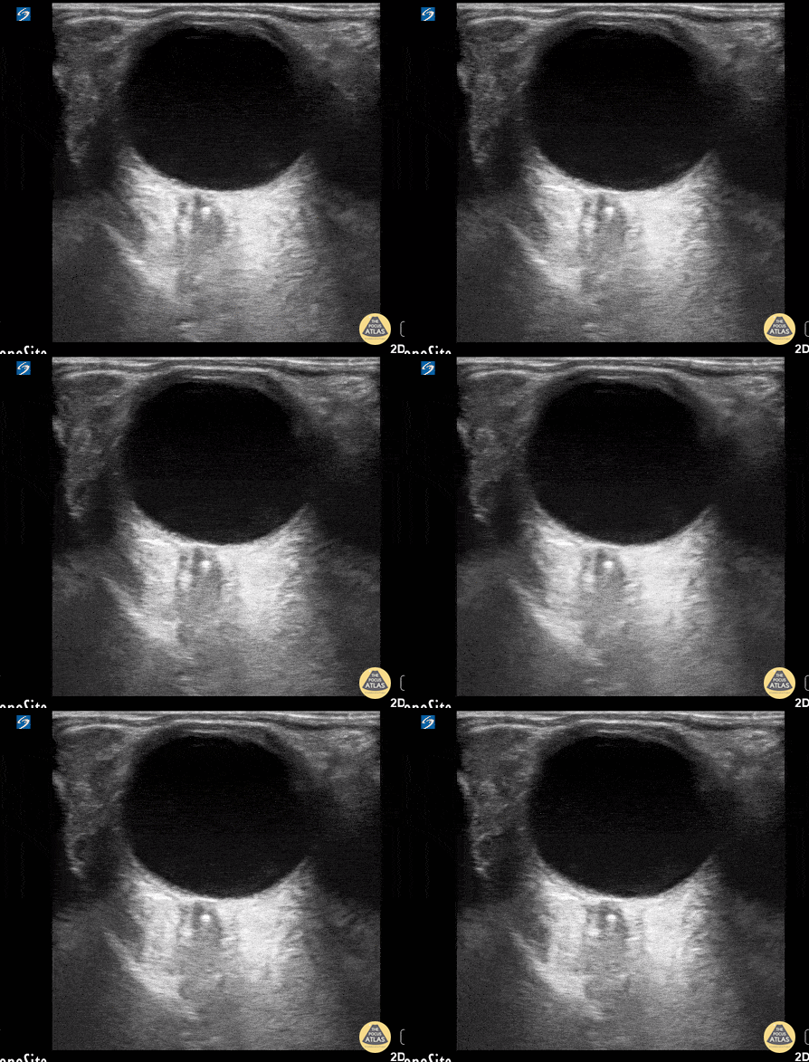
This was from a 52 year old male with a recent surgical history of vitreous detachment that became a retinal detachment. He presented to the emergency room for vision changes in same eye after an object fell and hit his head. Ocular ultrasound was performed at the bedside which revealed a new vitreous detachment. Important to note here that it is crucial to fan/tilt through the entire eye, making sure to visualize the optic nerve in order to differentiate between a vitreous and retinal detachment. In this case, notice how the lesion spotted in the chamber does not connect to the optic nerve, which is consistent with a vitreous detachment.
Dr. Christopher Paulo, DO, PGY-1
Riverside Regional Medical Center Emergency Medicine Program (Newport News, VA)

Central Retinal Artery Occlusion
This is from a 63 year old female who initially presented to the emergency department with vision loss over the last 24 hours. She reported bending over when she experienced complete vision loss from one of her eyes. Point of care ultrasound was performed, locating the optic nerve, but more interestingly a hyperechoic structure within the nerve (spot sign).
Overall, this is was suggestive of a central retinal artery occlusion. In this sort of situation, color doppler can also be utilized to assess for arterial versus venous occlusion.
Dr. Christopher Paulo, DO, PGY-1
Riverside Regional Medical Center Emergency Medicine Program (Newport News, VA)

Retrobulbar Hematoma
A 65-year-old patient presented with sudden-onset headache with associated right eye pain and diplopia. Physical exam also notable for an unsteady gait, exophthalmus, and impaired EOM including absent upward and medial gaze of right eye. Orbital POCUS revealed a retrobulbar hematoma causing external compression of the optic nerve.
Marco Garrone, Emergency Medicine Physician
@drmarcogarrone

Normal Eye Anatomy
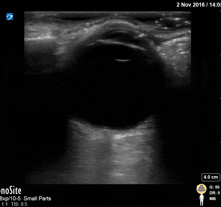
This image of the eye shows the following structures, from superficial to deep: eyelid, cornea (hyperechoic), anterior chamber (anechoic), iris (hyperechoic), lens (hyperechoic), vitreous (very large anechoic area), retina (flush with the posterior wall of the globe, not visible as a distinct structure under normal conditions), optic nerve sheath (hypoechoic structure extending midline perpendicularly from the globe at the bottom of the image).
Hannah Kopinksi and Dr. Lindsay Davis - NYU Emergency Medicine


