Orbital

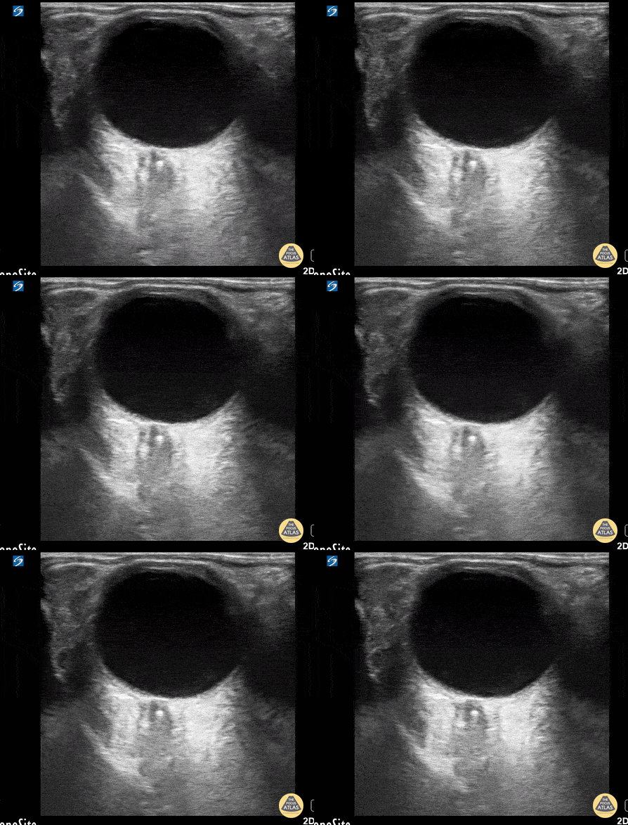
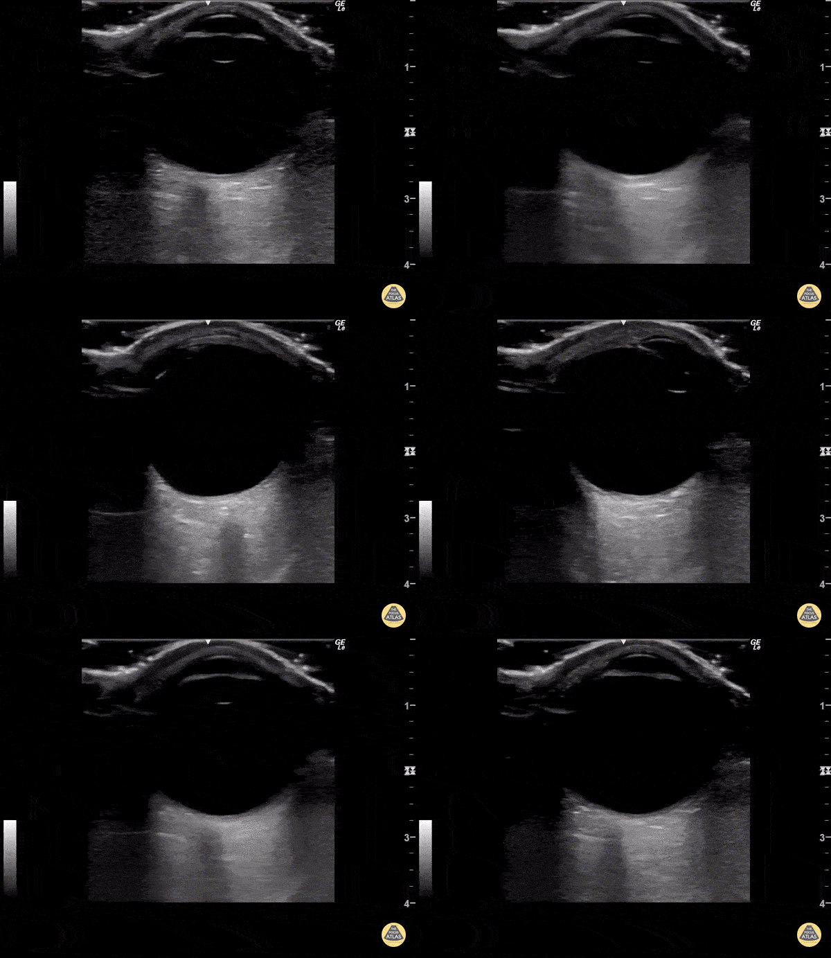
Prosthetic Ocular Lens Subluxation
This patient presented with decreased vision on a background of advanced macular degeneration. VA in the eye had decreased from 20/150 to 20/400 on presentation.
Interestingly the patient stated "I think my ocular lens has displaced."
Contributed by: Colin Bell, FRCPC, DPSPC

Papilledema From Optic Pathway Glioma
A 24-year old male presented to the ED with a six-month history of progressive total right vision loss. A bedside ocular ultrasound examination was performed that revealed an elevated optic disc with enlarged ONSD measuring 9.7mm, consistent with papilledema. An MRI of the head confirmed an enlarged intraconal portion of the right optic nerve, consistent with glioma.
Marko Lubardic, MS4; Tom Taugher, DO, PGY3; Michael Bernard, DO, PGY1; Central Michigan University Emergency Medicine Residency

Vitreous Detachment
This was from a 52 year old male with a recent surgical history of vitreous detachment that became a retinal detachment. He presented to the emergency room for vision changes in same eye after an object fell and hit his head. Ocular ultrasound was performed at the bedside which revealed a new vitreous detachment. Important to note here that it is crucial to fan/tilt through the entire eye, making sure to visualize the optic nerve in order to differentiate between a vitreous and retinal detachment. In this case, notice how the lesion spotted in the chamber does not connect to the optic nerve, which is consistent with a vitreous detachment.
Dr. Christopher Paulo, DO, PGY-1
Riverside Regional Medical Center Emergency Medicine Program (Newport News, VA)

Central Retinal Artery Occlusion
This is from a 63 year old female who initially presented to the emergency department with vision loss over the last 24 hours. She reported bending over when she experienced complete vision loss from one of her eyes. Point of care ultrasound was performed, locating the optic nerve, but more interestingly a hyperechoic structure within the nerve (spot sign).
Overall, this is was suggestive of a central retinal artery occlusion. In this sort of situation, color doppler can also be utilized to assess for arterial versus venous occlusion.
Dr. Christopher Paulo, DO, PGY-1
Riverside Regional Medical Center Emergency Medicine Program (Newport News, VA)

Retrobulbar Hematoma
Patient experienced ocular trauma 3 days prior to this exam, having been struck with a baseball to the eye. Notice the hypoechoic area within the retrobulbar space, consistent with a retrobulbar hematoma.
Tomasz Przednowek & Jereme Long @jplongest

Looking Around - Normal Exam
Normal ocular ultrasound over closed eyelid as patient moves eye medially and laterally.
Sukh Singh, MD


