Orbital

Intraoccular Lens Subluxation
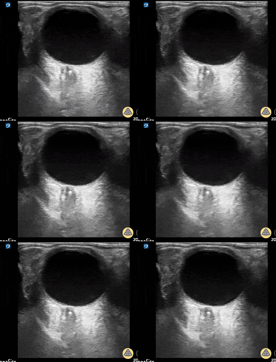
A 50 year old male with a prior history of bilateral intraoccular lens (IOL) transplants presented to our ED with sudden onset foggy vision in his right eye while getting out of the shower. He was unable to participate in visual acuity due to the extent of his blurred. POCUS demonstrated “iridodonesis” and a provisional diagnosis of IOL subluxation/dislocation was made. Ophthalmology was consulted and confirmed the diagnosis.
Dr. Piaseczny, PGY4 Emergency Medicine, Queen's University, Kingston, Ontario, Canada

Vitreous Detachment
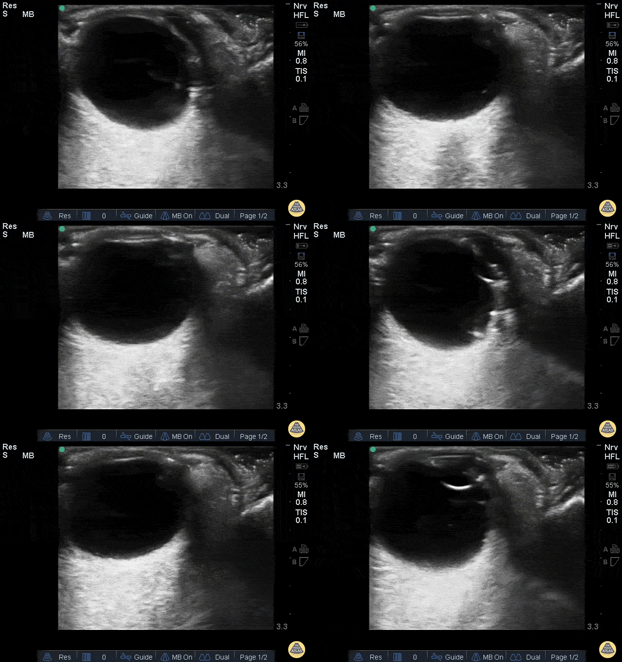
This was from a 52 year old male with a recent surgical history of vitreous detachment that became a retinal detachment. He presented to the emergency room for vision changes in same eye after an object fell and hit his head. Ocular ultrasound was performed at the bedside which revealed a new vitreous detachment. Important to note here that it is crucial to fan/tilt through the entire eye, making sure to visualize the optic nerve in order to differentiate between a vitreous and retinal detachment. In this case, notice how the lesion spotted in the chamber does not connect to the optic nerve, which is consistent with a vitreous detachment.
Dr. Christopher Paulo, DO, PGY-1
Riverside Regional Medical Center Emergency Medicine Program (Newport News, VA)

Central Retinal Artery Occlusion
This is from a 63 year old female who initially presented to the emergency department with vision loss over the last 24 hours. She reported bending over when she experienced complete vision loss from one of her eyes. Point of care ultrasound was performed, locating the optic nerve, but more interestingly a hyperechoic structure within the nerve (spot sign).
Overall, this is was suggestive of a central retinal artery occlusion. In this sort of situation, color doppler can also be utilized to assess for arterial versus venous occlusion.
Dr. Christopher Paulo, DO, PGY-1
Riverside Regional Medical Center Emergency Medicine Program (Newport News, VA)

Retinal Detachment
Retinal Detachment.
Francisco Norman

Vitreous Hemorrhage due to Proliferative Diabetic Retinopathy
A 70-year-old man presented with loss of vision to his right eye (hand motion and light perception). He takes warfarin and has diabetes. Monocular vision loss triggered a POCUS that revealed a hazy echogenic substance (consistent with blood) seen best when patient activates extra-ocular muscle movements. Also note the associated bright linear structure within one portion of the bleed, the hyaloid membrane. This patient was subsequently assessed by the Eye Hospital and diagnosed with vitreous hemorrhage due to proliferative diabetic retinopathy.
Peter Cheng

Vitreous Hemorrhage
A 71-year-old male with a history of hypertension and diabetes presented to the ED reporting gradual vision loss in his right eye over a 3-day period. Physical exam was notable for a 3 mm and nonreactive pupil with ipsilateral visual acuity reduced to patient only being able to detect movement. POCUS revealed swirling echodensity within the right eye, most appreciable with extraocular muscle movements. Ophthalmology subsequently confirmed our suspected diagnosis of vitreous hemorrhage.
Richard Cunningham, MD @HappyDays_EM
Maricopa Medical Center

Endogenous Endophthalmitis
A middle aged female with ESRD and longstanding percutaneous HD catheter with recent MSSA bacteremia was admitted with septic shock. She subsequently developed subacute bilateral visual loss (OS > OD). Clinical suspicion of endogenous endophthalmitis was initially supported by POCUS notable for heterogenous intraoccular material within vitreous. She was immediately started in intravitreal antibiotics in addition to previously initiated systemic antibiotics. Diagnosis of endogenous endophthalmitis was subsequently confirmed by vitrectomy.
Tessa W. Damm, DO
Intensivist, Critical Care Medicine & Neurocritical Care.
Wisconsin, USA.
@DrDamm

Vitreous vs. Retinal Detachment
A 44 year old man came to ED with a shimmering effect in his left eye and unilateral temporary painless visual disturbance. He had previously been treated for a retinal tear.
Ocular PoCUS shows a frond-like linear structure lifting away from the posterior surface of the globe.
Posterior vitreous detachment (PVD) was suspected while retinal detachment (RD) was also considered. PVD was confirmed by the specialist team and the patient was treated conservatively.
Dr. Cian McDermott, Emergency Physician - University Hospital Geelong, Australia


