
Peds-Gastrointestinal

Pediatric Acute Appendicitis
Teenage male presenting with abdominal pain worsening over 24 hours, diagnosed with acute appendicitis via POCUS.
The clip shows a circular structure which measures at 6.5 mm transversely, representing an acutely inflamed appendix with surrounding anechoic free fluid.
POCUS Acute Appendicitis: noncompressible, diameter >6mm, single wall >3mm are direct signs of appendicitis. Use of POCUS for diagnosis of acute appendicits significantly reduces cost and radiation exposure for patients without sacrificing diagnostic accuracy.
Contributor: C. Malcolm Roberson, MD. @ProjectUltraEM
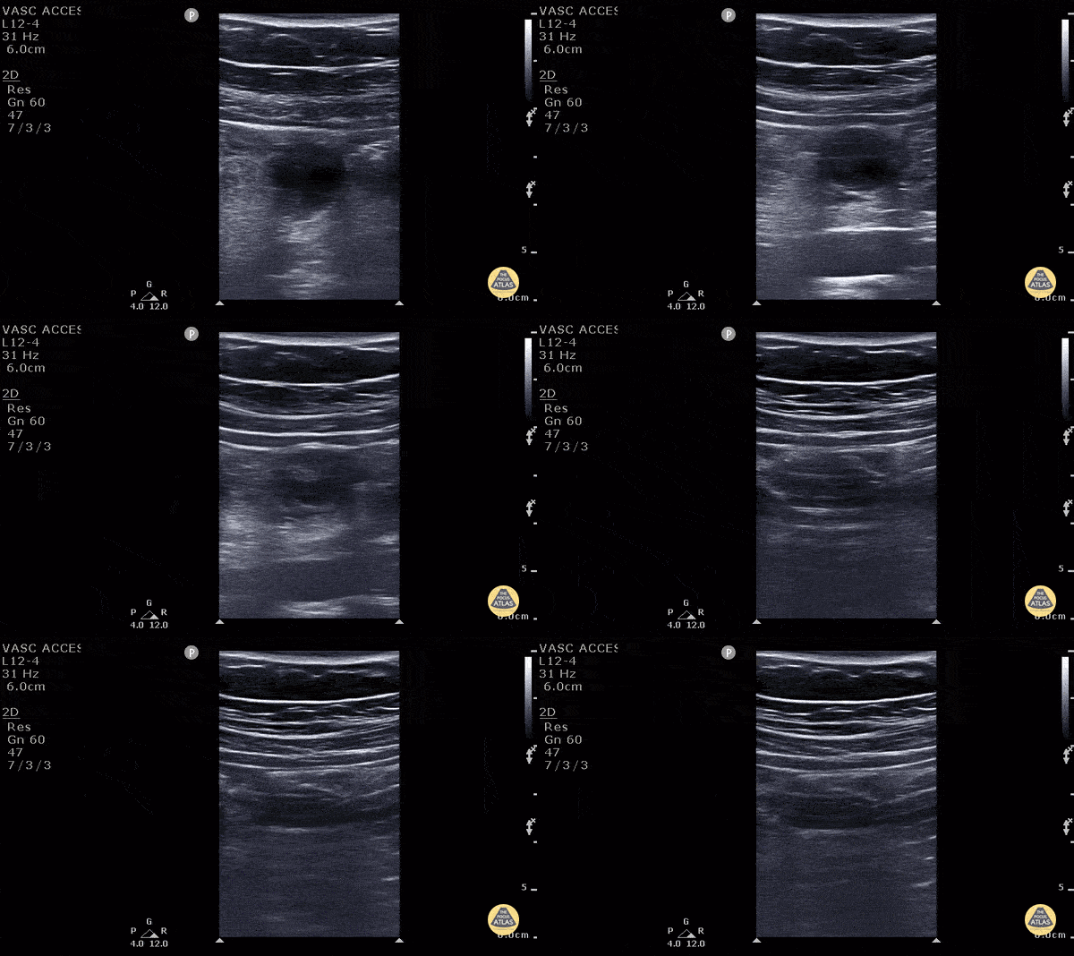
Appendicitis (2/2) - Transverse
11 y/o M presented with 1 day of periumbilical pain that migrated to the right lower quadrant with nausea, vomiting and anorexia. Ultrasound with high frequency linear probe demonstrates an enlarged appendix with an external diameter of 1.32 cm with trace free fluid posteriorly, as well as a fecalith at the proximal end of the appendix. Surgery was consulted who requested a formal US which was non-diagnostic. Surgery took the patient anyway, and MRI confirmed our findings (including the large diameter).
Diagnostic criteria for appendicitis: a non-compressible, aperistaltic, blind ended structure >6mm diameter. Visualizing free fluid, tenderness in that area, and visualizing a fecalith can also add to the diagnosis.
See the evidence atlas for more info about POCUS diagnosing appendicitis but when visualizing a diagnostic appendicitis, it carries a positive likelihood ratio of 9.24. PMID: 28214369
Jackie Chiou MS4, Dr. Matthew Riscinti and Dr. Tian Liang - Kings County Emergency Medicine

Pyloric Stenosis - Antral Nipple Sign
3 week old with projectile vomiting, POCUS showed positive astral nipple sign which is a highly specific finding for pyloric stenosis - the redundant pyloric mucosa protrudes into the gastric antrum. The measurements show increased pyloric muscle thickness (>3mm) and increased pyloric longitudinal measurement (>15 - 17 mm)
Measurements can be remembered using "Pi Rule"
- Pyloric muscle thickness, i.e. diameter of a single muscular wall on a transverse image >3 mm
- Pyloric transverse diameter >14 mm
- Pyloric longitudinal measurement >15 - 17 mm
Contributed by:
Dimitri Livshits DO, Ultrasound Fellow; Jane Belyavskaya MD, Ultrasound Fellow; Chris Hanuscin MD, Ultrasound Division Director (Kings County/SUNY Downstate)
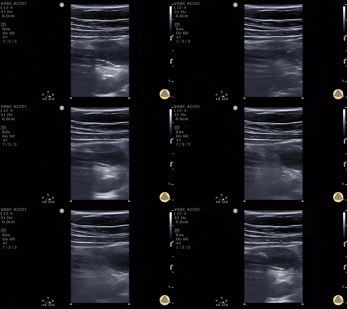
Appendicitis (1/2) - Longitudinal
11 y/o M presented with 1 day of periumbilical pain that migrated to the right lower quadrant with nausea, vomiting and anorexia. Ultrasound with high frequency linear probe demonstrates an enlarged appendix with an external diameter of 1.32 cm with trace free fluid posteriorly, as well as a fecalith at the proximal end of the appendix. Surgery was consulted who requested a formal US which was non-diagnostic. Surgery took the patient anyway, and MRI confirmed our findings (including the large diameter).
Diagnostic criteria for appendicitis: a non-compressible, aperistaltic, blind ended structure >6mm diameter. Visualizing free fluid, tenderness in that area, and visualizing a fecalith can also add to the diagnosis.
See the evidence atlas for more info about POCUS diagnosing appendicitis but when visualizing a diagnostic appendicitis, it carries a positive likelihood ratio of 9.24. PMID: 28214369
Jackie Chiou MS4, Dr. Matthew Riscinti and Dr. Tian Liang - Kings County Emergency Medicine

Pyloric Stenosis - Antral Nipple Sign with Measurements
3 week old with projectile vomiting, POCUS showed positive astral nipple sign which is a highly specific finding for pyloric stenosis - the redundant pyloric mucosa protrudes into the gastric antrum. The measurements show increased pyloric muscle thickness (>3mm) and increased pyloric longitudinal measurement (>15 - 17 mm)
Measurements can be remembered using "Pi Rule"
- Pyloric muscle thickness, i.e. diameter of a single muscular wall on a transverse image >3 mm
- Pyloric transverse diameter >14 mm
- Pyloric longitudinal measurement >15 - 17 mm
Contributed by:
Dimitri Livshits DO, Ultrasound Fellow; Jane Belyavskaya MD, Ultrasound Fellow; Chris Hanuscin MD, Ultrasound Division Director (Kings County/SUNY Downstate)
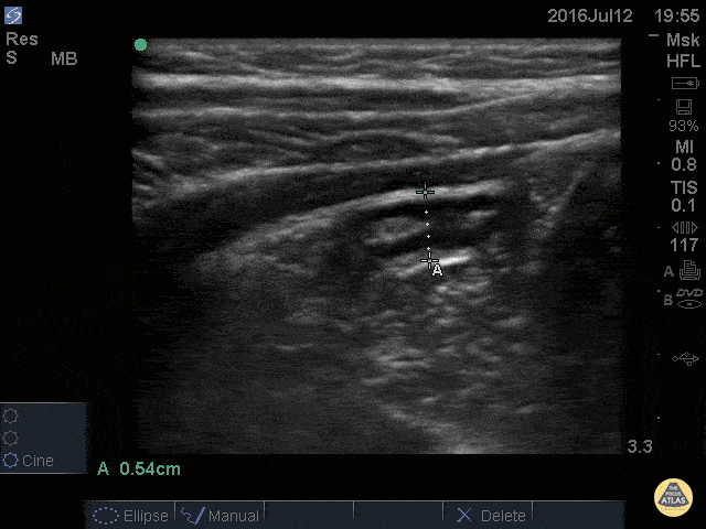
Normal Appendix - Cross Section Measured
14 y/o M with nausea vomiting and and RLQ pain. POCUS visualized a normal appendix is seen. A normal appendix is identified by a blind-ending tubular structure that is <6mm diameter measured from outer wall to outer wall (although 6mm-7mm has also been described). This patient’s appendix was measure to be 5.4mm.
Dr. Sathya Subramaniam - Children’s Hospital of Philadelphia
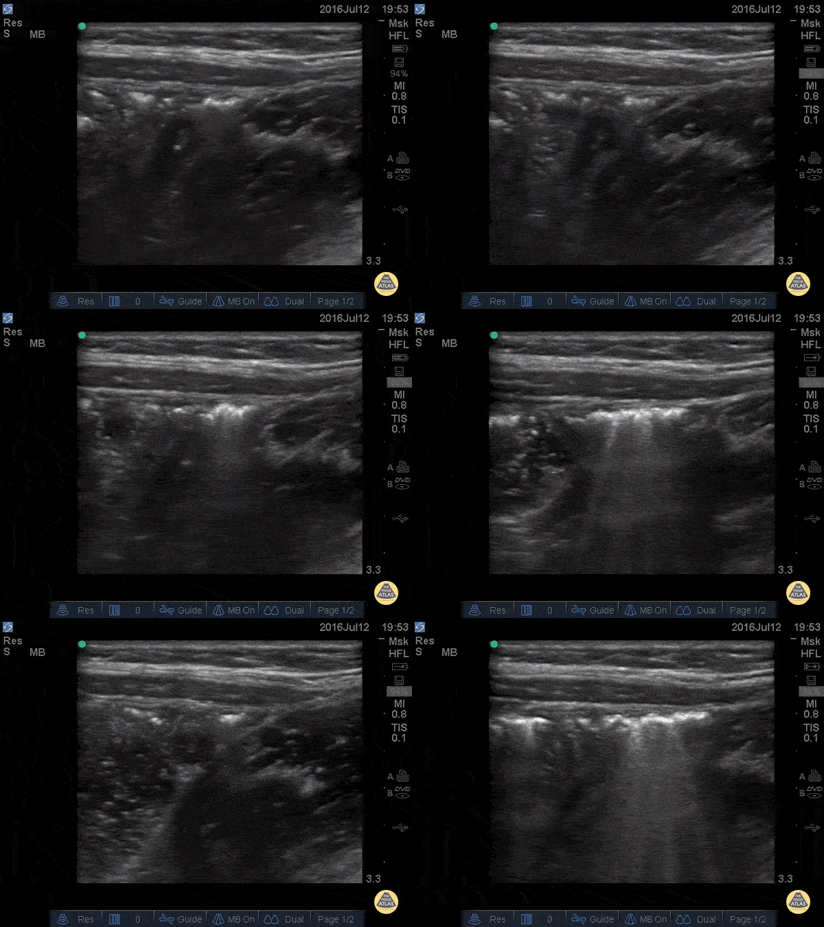
Normal Appendix - Cross Section
14 y/o M with nausea vomiting and and RLQ pain. POCUS visualized a normal appendix is seen. A normal appendix is identified by a blind-ending tubular structure that is <6mm diameter measured from outer wall to outer wall (although 6mm-7mm has also been described). This patient’s appendix was measure to be 5.4mm (see still image).
Dr. Sathya Subramaniam - Children’s Hospital of Philadelphia
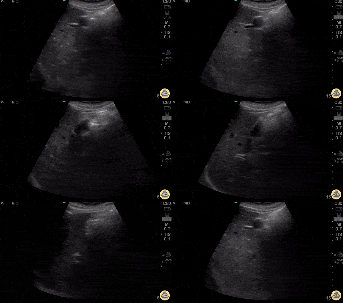
Splenic Sequestration
11 y/o F with PMH of sickle cell disease (ss) presents with 2 hours of tactile temperature, chest pain, and vague abdominal pain. Exam demonstrates normal vitals (T99.3) distended abdomen, nonspecific mild tenderness to palpation and enlarged spleen tip to the umbilicus. Ultrasound was used to confirm the size spanning nearly >15cm in length with heterogeneous echogenicity throughout the spleen consistent with splenic sequestration syndrome.
Dr. Praneetha Chaganti and Dr. Eddie Rodriguez - Kings County Emergency Medicine


