
Peds-Gastrointestinal

Appendicolith
Transverse view of the appendix showing the appendix with intraluminal appendicolith.
Contributor: Maher M. Abulfaraj, MD, @mahermabulfaraj

Ascites
Ascites with floating bowel and questionable to-and-fro peristalsis.
Contributor: Peter Gutierrez, MD, FAAP, Emory University School of Medicine/Children's Healthcare of Atlanta, @pocuspete

Pseudokidney Sign - Intussusception
23 month old with ileocolic intussusception. Pseudokidney sign seen here due to oblique orientation of the linear transducer.
Contributor: Antonio Riera, MD

Esophageal FB
5 yo male presents with swallowed decorative marble. no difficulty breathing. unable to handle secretions. POCUS shows an esophageal FB that was later removed by surgery.
Contributor: Paul Khalil, MD Nicklaus Children's Hospital, @khalil3paul

Neuroblastoma
18 mo F sent from PMD for mass in the RUQ. POCUS shows extra renal mass consistent with neuroblastoma that was later confirmed by pathology.
Contributor: Paul Khalil, MD Nicklaus Children's Hospital
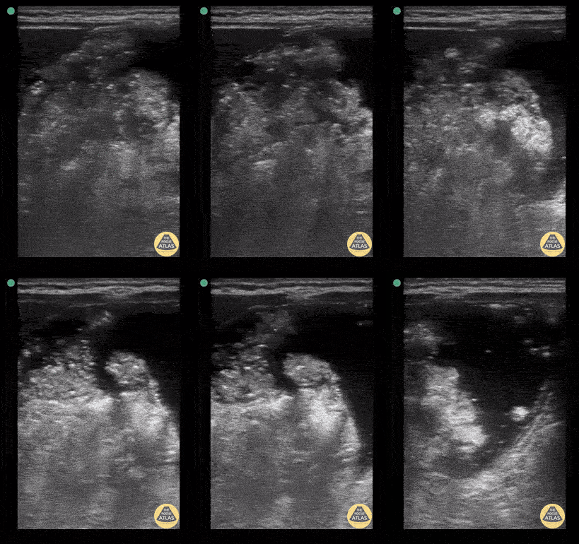
Full stomach 1
Gastric content eval for patient undergoing procedural sedation. With the patient in the right lateral decubitus position the linear probe is placed in a subxiphoid sagittal axis with the probe marker towards the head. The stomach is seen immediately caudal to the liver. The class 5 layer bowel wall of the stomach can be seen containing large volume, mixed echogenicity content.
Contributor: Matthew Moake, MD PhD
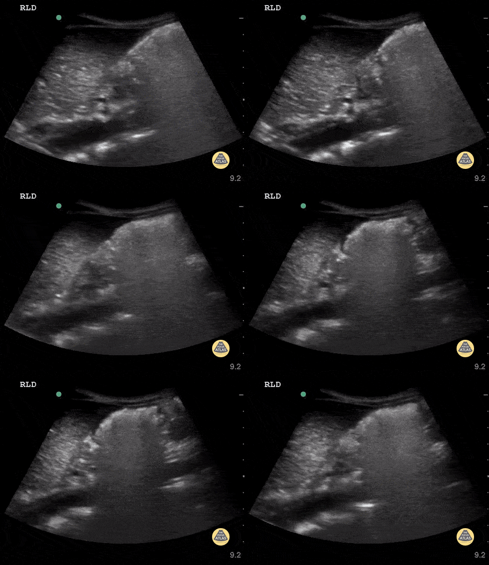
Full stomach 2
Ultrasound used to assess gastric content prior to procedural sedation. Sagittal view in the epigastric region using the curvilinear probe with the patient in the right lateral decubitus position. Probe indicator cephalad. The gastric antrum is seen in the upper right with heavily air-admixed content with dirty shadow obscuring deeper content. The liver is seen in the upper left and the aorta in the deep field.
Contributor: Matthew Moake, MD PhD
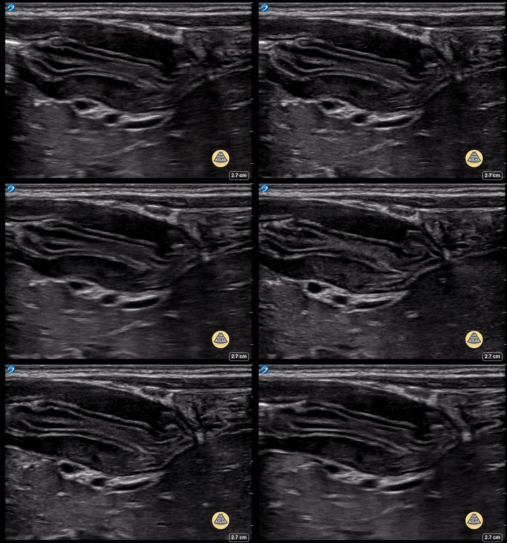
Pyloric Stenosis 1
Roughly 1m male with projectile emesis. POCUS demonstrated elongated and thickened pylorus with absence of trans-pyloric flow.
Contributor: Matthew Moake, MD PhD
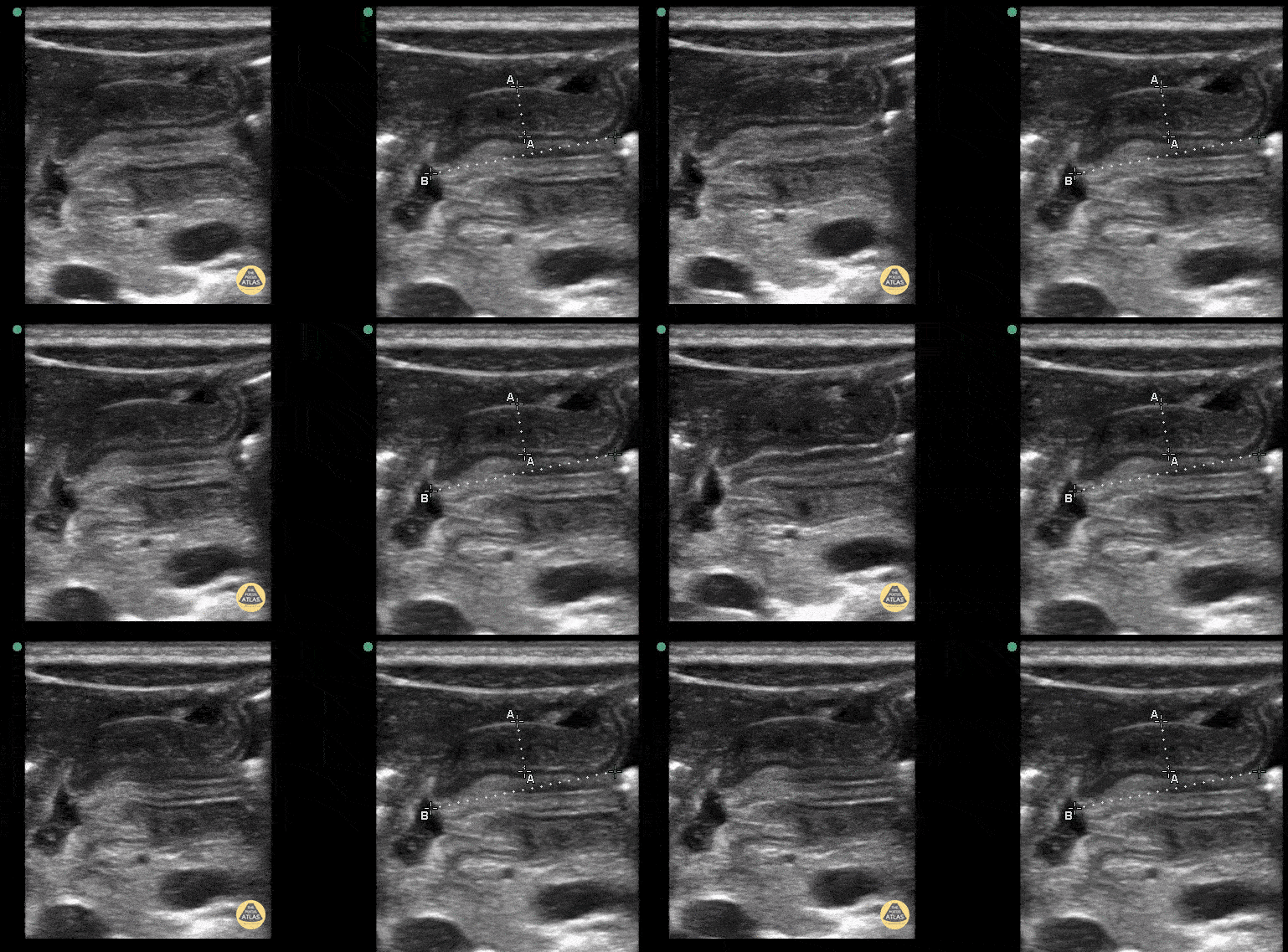
Pyloric Stenosis 2 (1/2)
2+ weeks projectile vomiting. Extreme lab abnormalities including Cl 67. CG4: 7.57/67/34/60, base excess >30, lactate 3.23. The pyloric channel is elongated, thickened, and has an absence of trans-pyloric flow.
Contributor: Matthew Moake, MD PhD

Pyloric Stenosis 2 (2/2)
2+ weeks projectile vomiting. Extreme lab abnormalities including Cl 67. CG4: 7.57/67/34/60, base excess >30, lactate 3.23. The pyloric channel is elongated, thickened, and has an absence of trans-pyloric flow.
Contributor: Matthew Moake, MD PhD
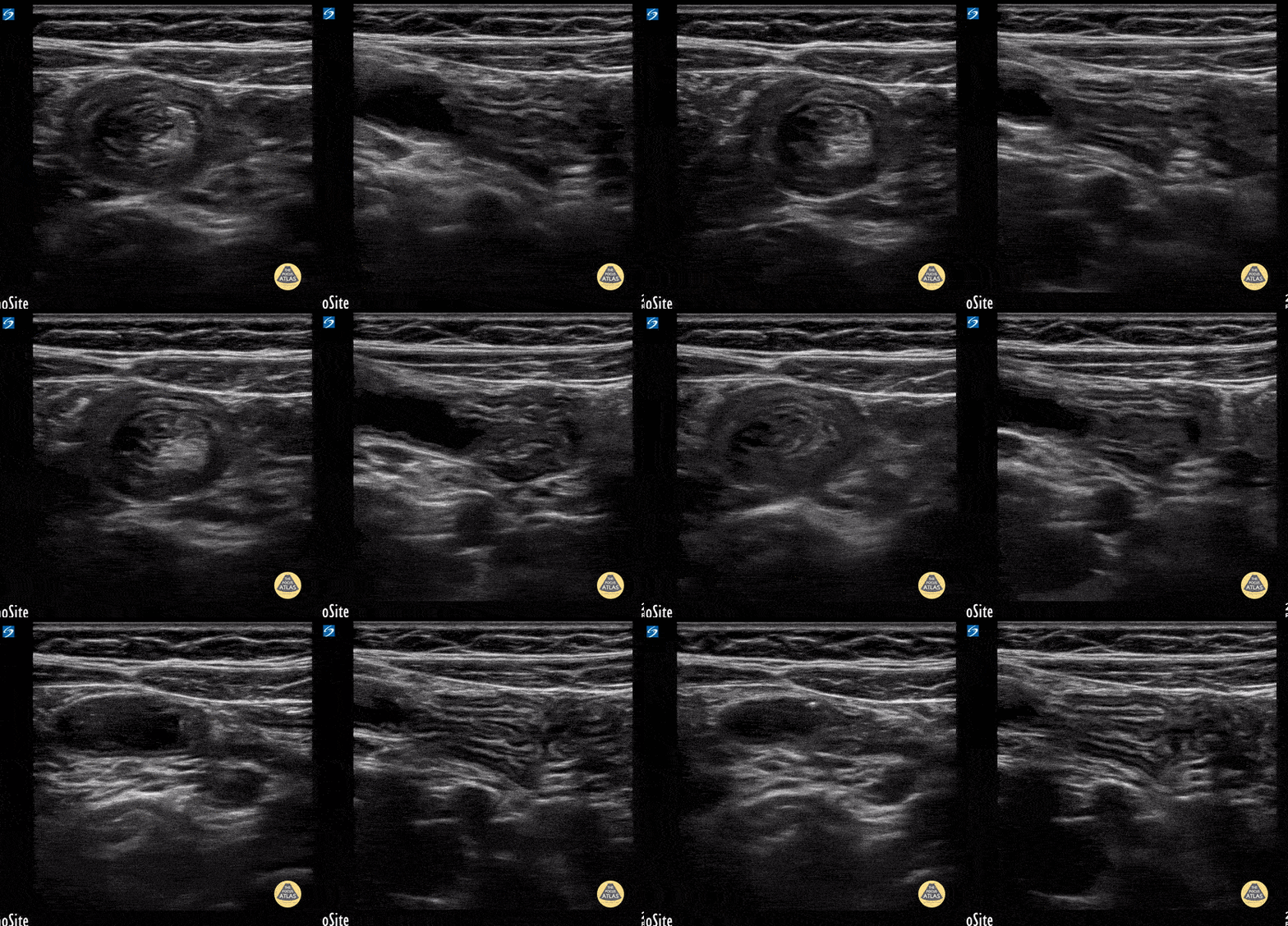
Ileo-ileal intussusception
7y female with N/V and tactile fever. Benign abdominal exam. Note the small size of the intuss. In long axis you can easily track the bowel wall as it folds into itself and see it is slowly sliding in and out a small bit.
Contributor: Matthew Moake, MD PhD
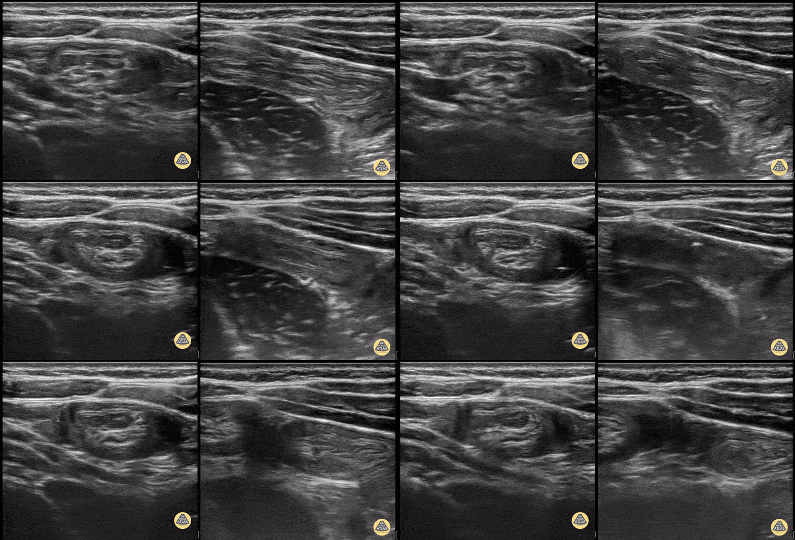
Ileo-ileal Intussuception 2
Toddler with colicky abdominal pain. LUQ with ileo-ileal intuss. Note the smaller size and active peristalsis of the intuss.
Contributor: Matthew Moake, MD PhD
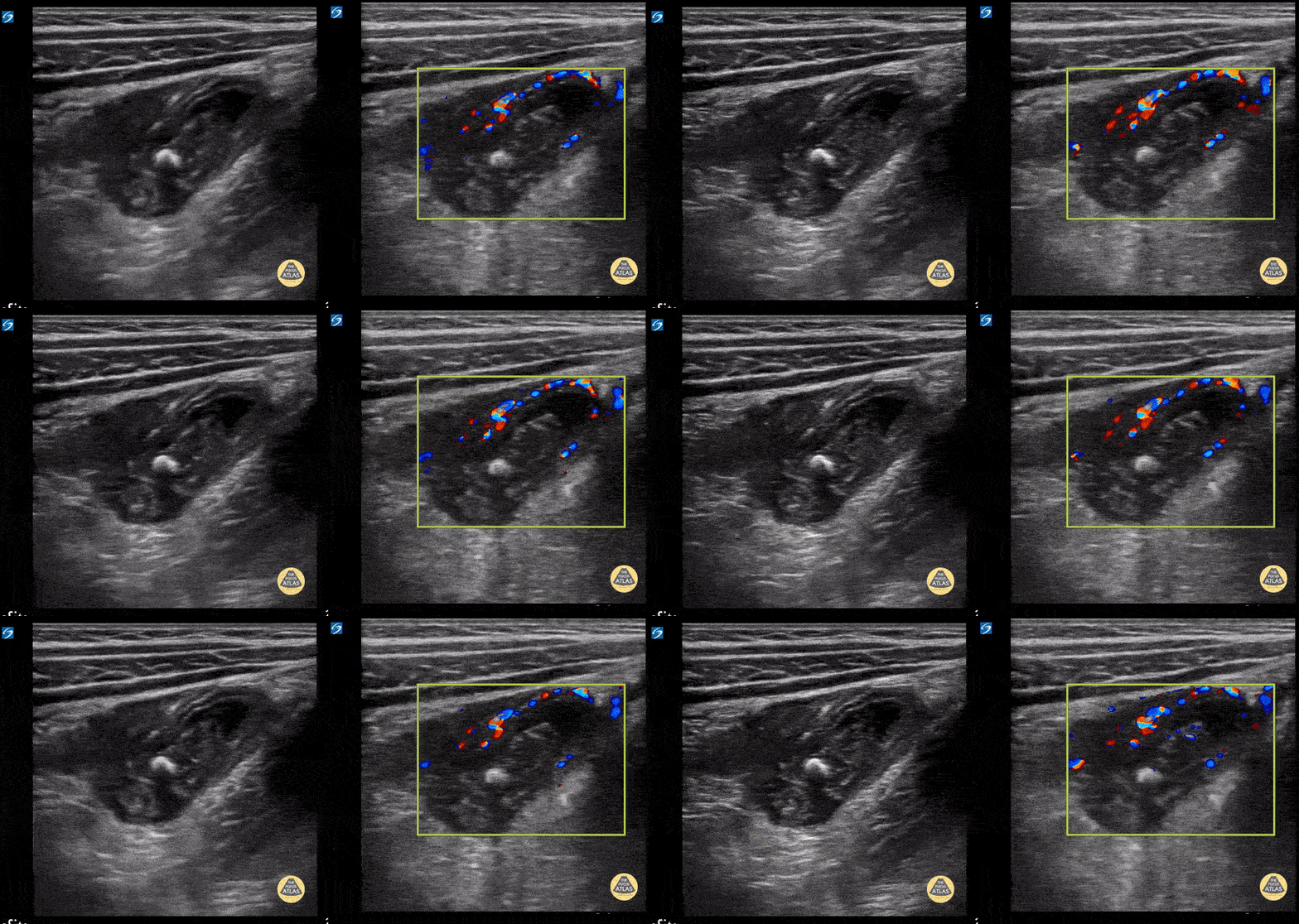
Ruptured Appendicitis
3y female with 3d RLQ abd pain, emesis, fever. WBC 8.5 with 73% PMN. CRP 9. POCUS with enlarged appendix with loss of bowel wall architecture, surrounding early fluid collection, fecolith, and mural hyperemia.
Contributor: Matthew Moake, MD PhD
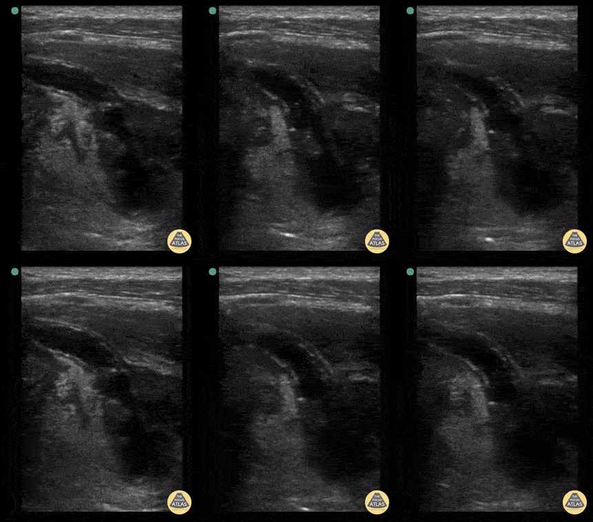
Ruptured Appendicitis 2
6y female with 2d abd pain, fever, nauesea, dysuria. OSH WBC 17.5, pyuria. Long axis view of the appendix draping down into the pelvis. Note how the regular bowel wall architecture is progressively obliterated as the appendix tracks distally into the pelvis, ending in full perforation with early abscess formation and surrounding hyperechoic fat.
Contributor: Matthew Moake, MD PhD
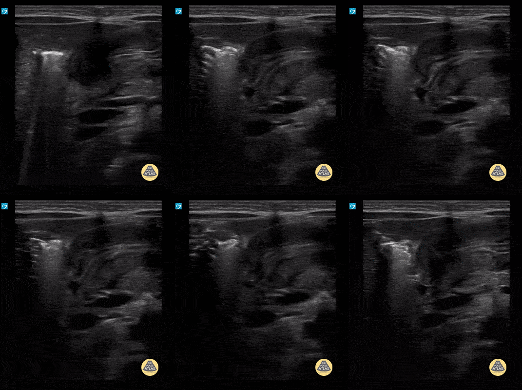
Pyloric Stenosis with and without measurements (1/4)
5 wk old male with projectile vomiting. Clips show hypertrophic pyloric stenosis in long axis.
Contributor: Paul Khalil, MD Nicklaus Children's Hospital @khalil3paul
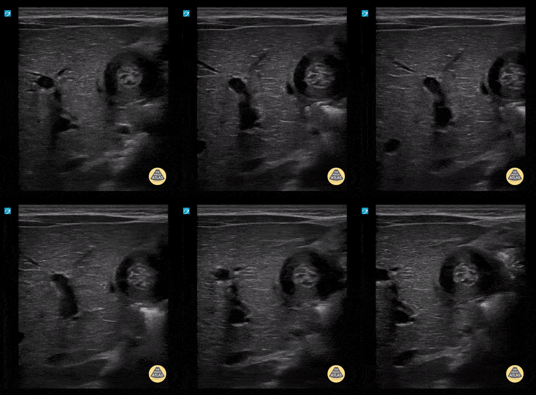
Pyloric Stenosis with and without measurements (2/4)
5 wk old male with projectile vomiting. Clips show hypertrophic pyloric stenosis in short axis.
Contributor: Paul Khalil, MD Nicklaus Children's Hospital @khalil3paul
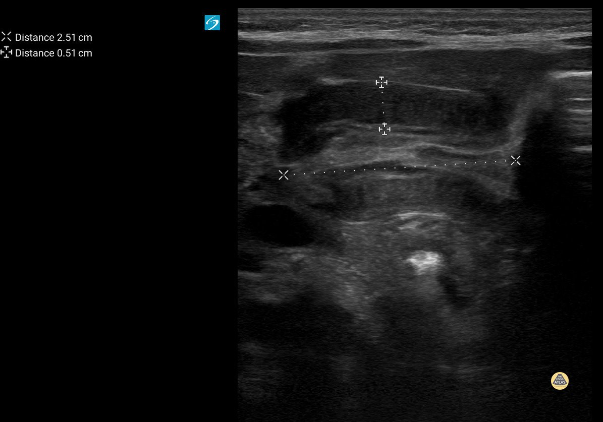
Pyloric Stenosis with and without measurements (3/4)
5 wk old male with projectile vomiting. Clips show hypertrophic pyloric stenosis in long axis.
Contributor: Paul Khalil, MD Nicklaus Children's Hospital @khalil3paul

Pyloric Stenosis with and without measurements (4/4)
5 wk old male with projectile vomiting. Clips show hypertrophic pyloric stenosis in short axis.
Contributor: Paul Khalil, MD Nicklaus Children's Hospital @khalil3paul
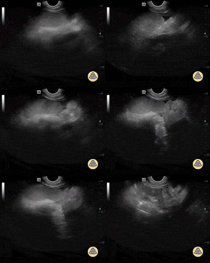
Dengue Ascites
4-year-old boy with severe dengue with free fluid in the abdominal cavity.
Contributor: Mg. Andres Silva Horna, Hospital Cayetano Heredia Piura-Peru
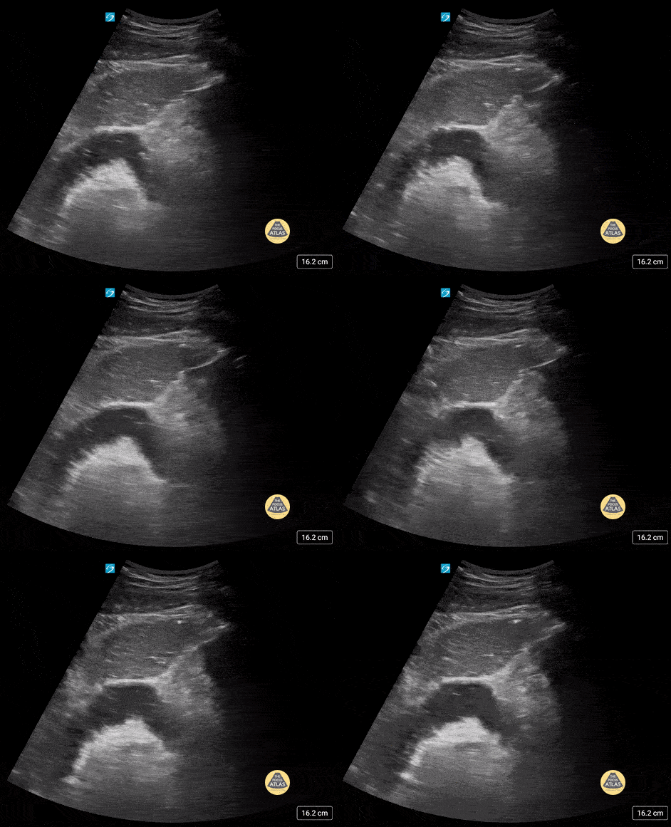
SBO
17 yo male with history of abdominal surgeries (g-tube and fundoplication) and chronic constipation who comes in with lower abdominal pain. POCUS shows stool to and fro (tanga sign).
Contributor: Paul Khalil, MD Nicklaus Children's Hospital @khalil3paul

Neurofibroma Abdominal Mass
3 yo presents with enlarging abdomen and mass palpated. A large abdominal mass was seen on POCUS. The kidney is seen directly below the mass in the image.
Contributor: Kathryn Pade, MD, Rady Children's Hospital San Diego
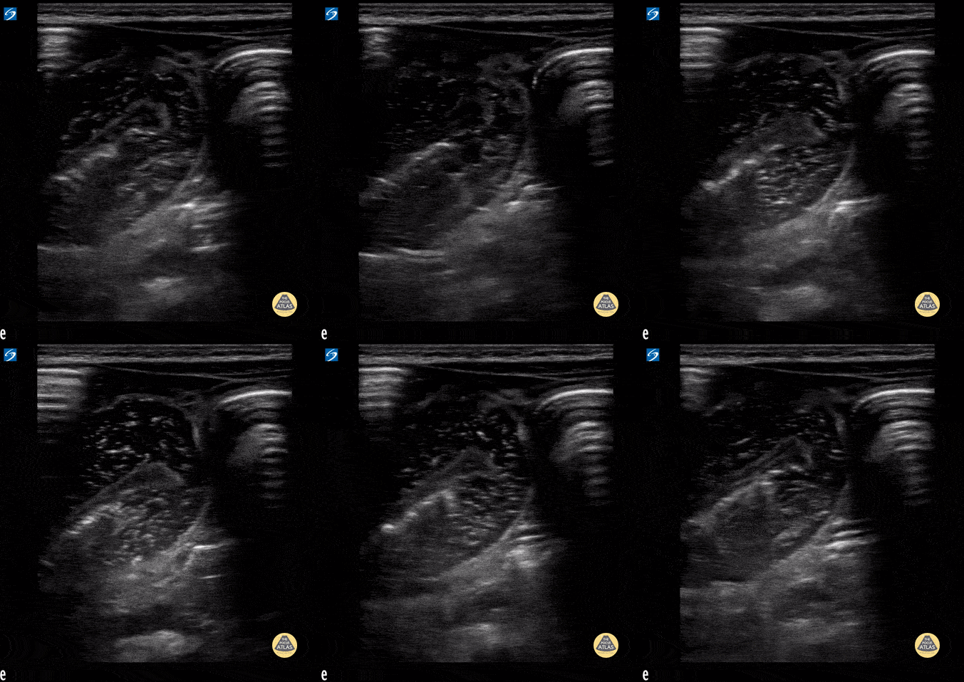
SBO
Small bowel obstruction with to and fro peristalsis visualized.
Contributor: Peter Gutierrez, MD FAAP FACEP; Children's Healthcare of Atlanta; @pocuspete

Ileocolic Intussusception
Ileocolic intussusception measuring 4.4cm.
Contributor: Peter Gutierrez, MD FAAP FACEP; Children's Healthcare of Atlanta; @pocuspete
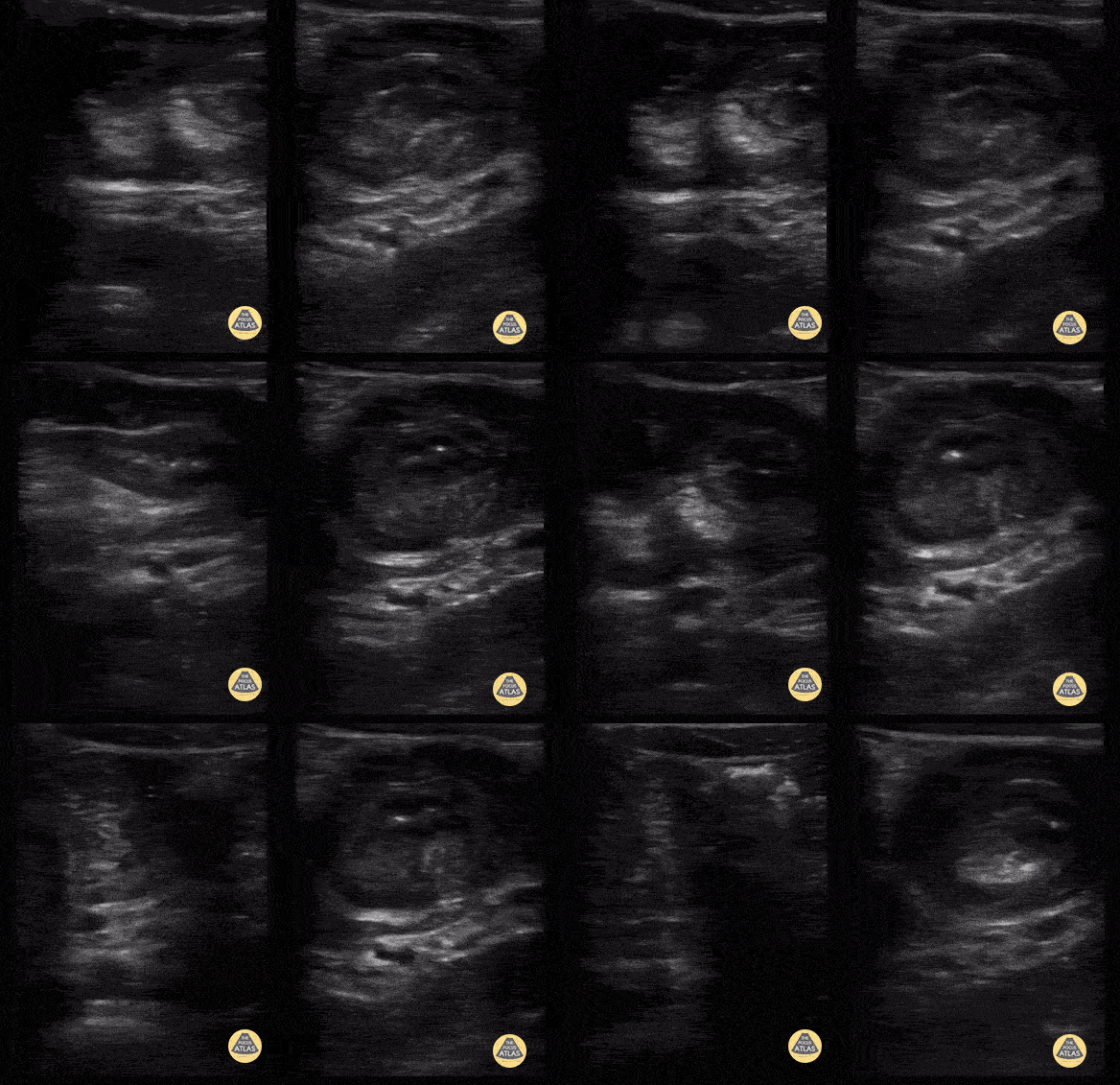
Ileocolic LUQ intussusception - Longitudinal & Transverse
3 yo male presented with an episode of abdominal pain and bilious emesis in the morning. He was noted to be sleepy all day at home and passed two bloody diarrheal stools prior to arrival. Point-of-care ultrasound demonstrates ileocolic intussusception in the left upper quadrant. Transverse view demonstrate a "target sign" and longitudinal view demonstrates "sandwich sign" consistent with the diagnosis.
Contributor: Megan Musisca, MD, Boston Children's Hospital
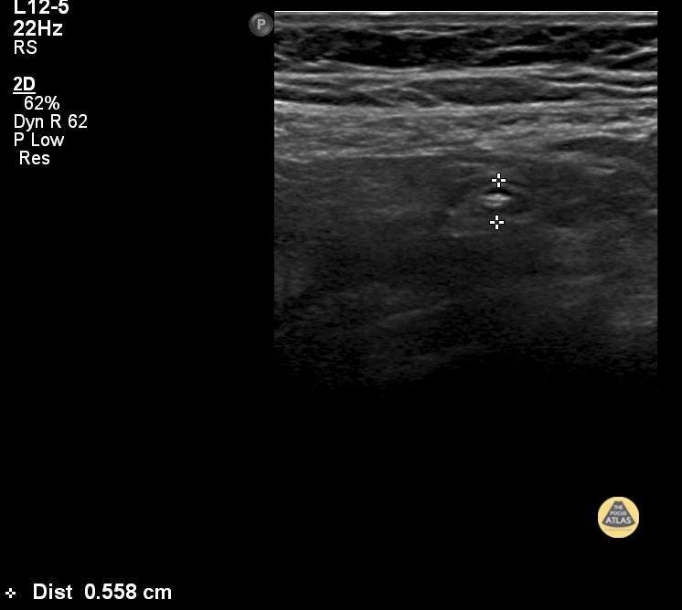
Appendicitis (1/3)
"Fever Vomiting and Abdominal pain 9yr old
Tenderness @ McBurney’s point
USG : Aperistaltic Dilated with probe tenderness in right iliac region Diameter 6mm plus"
Contributor: Dr Vanitha Jagannath

Appendicitis (2/3)
"Fever Vomiting and Abdominal pain 9yr old
Tenderness @ McBurney’s point
USG : Aperistaltic Dilated with probe tenderness in right iliac region Diameter 6mm plus"
Contributor: Dr Vanitha Jagannath
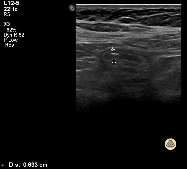
Appendicitis (3/3)
"Fever Vomiting and Abdominal pain 9yr old
Tenderness @ McBurney’s point
USG : Aperistaltic Dilated with probe tenderness in right iliac region Diameter 6mm plus"
Contributor: Dr Vanitha Jagannath
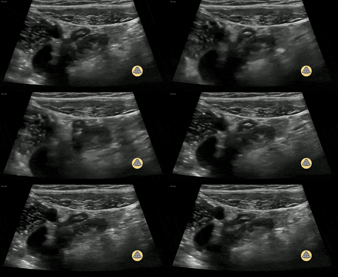
Normal Appendix 1 (1/3)
10 y/o with abdominal pain. Normal appendix identified medial to the iliac vessels. Please see other image in series for doppler.
Contributor: Elena Chen, MD
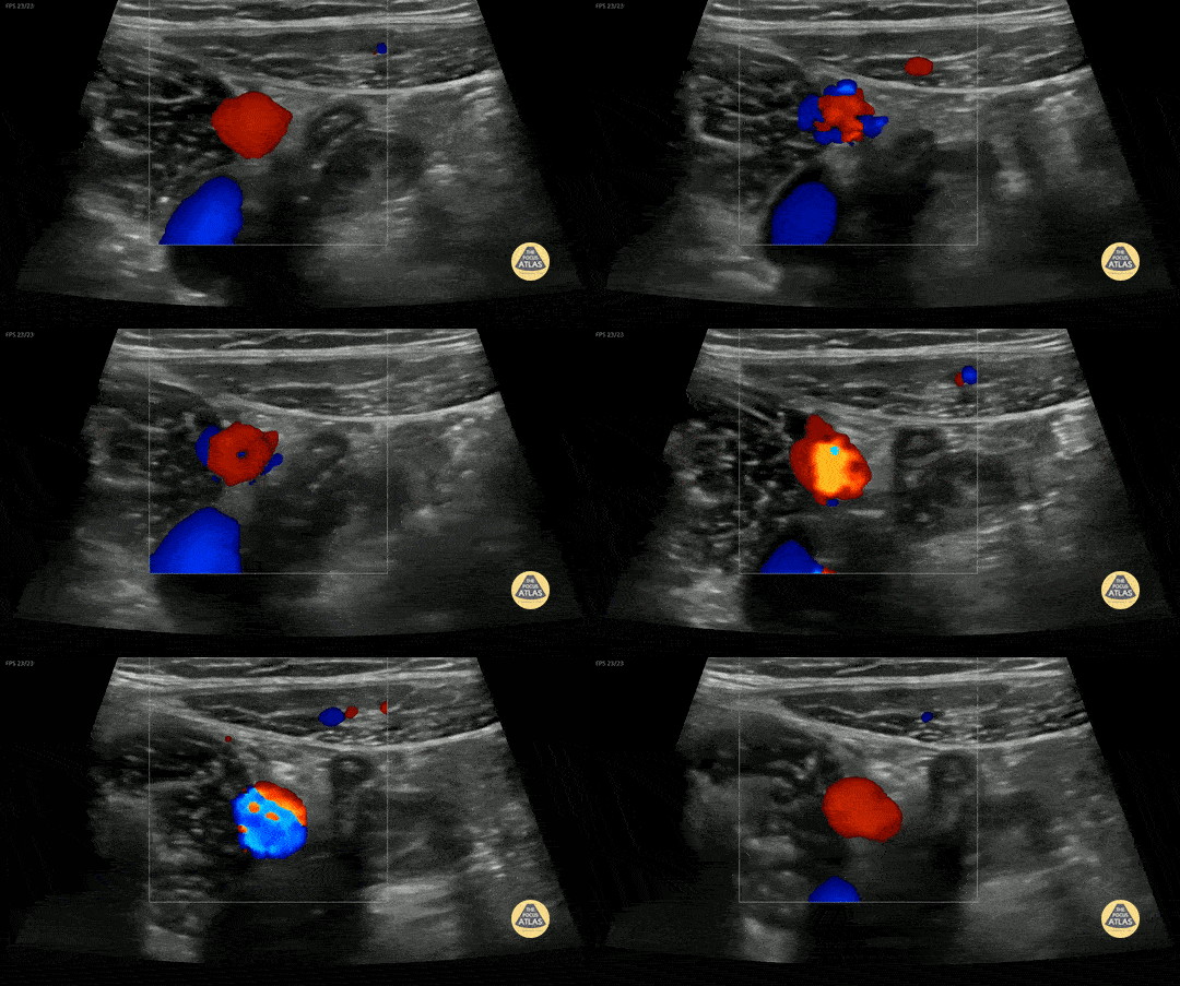
Normal Appendix 1 (2/3)
10 y/o with abdominal pain. Normal appendix identified medial to the iliac vessels.
Contributor: Elena Chen, MD

Normal Appendix 1 (3/3)
10 y/o with abdominal pain. Normal appendix identified medial to the iliac vessels.
Contributor: Elena Chen, MD

Normal Appendix 2 (1/2)
7 y/o F with abdominal pain. Normal appendix identified.
Contributor: Russ Horowitz, MD, Lurie Children's Hospital

Normal Appendix 2 (2/2)
7 y/o F with abdominal pain. Normal appendix identified.
Outer wall measured 0.48cm in long, 0.46cm in short, and 0.51cm again in a short axis.
Contributor: Russ Horowitz, MD, Lurie Children's Hospital


