
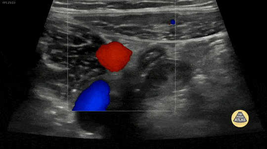
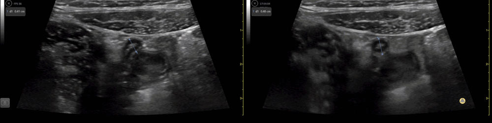
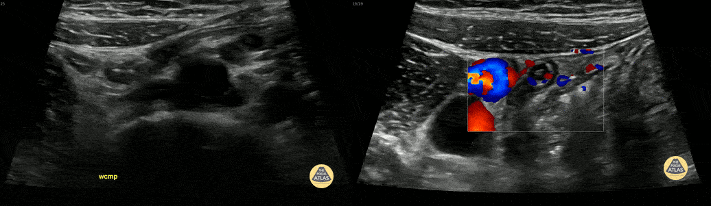
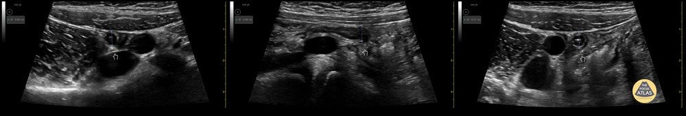
Peds-Gastrointestinal

Full stomach 1
Gastric content eval for patient undergoing procedural sedation. With the patient in the right lateral decubitus position the linear probe is placed in a subxiphoid sagittal axis with the probe marker towards the head. The stomach is seen immediately caudal to the liver. The class 5 layer bowel wall of the stomach can be seen containing large volume, mixed echogenicity content.
Contributor: Matthew Moake, MD PhD

Full stomach 2
Ultrasound used to assess gastric content prior to procedural sedation. Sagittal view in the epigastric region using the curvilinear probe with the patient in the right lateral decubitus position. Probe indicator cephalad. The gastric antrum is seen in the upper right with heavily air-admixed content with dirty shadow obscuring deeper content. The liver is seen in the upper left and the aorta in the deep field.
Contributor: Matthew Moake, MD PhD

Normal Appendix 1 (1/3)
10 y/o with abdominal pain. Normal appendix identified medial to the iliac vessels. Please see other image in series for doppler.
Contributor: Elena Chen, MD

Normal Appendix 1 (2/3)
10 y/o with abdominal pain. Normal appendix identified medial to the iliac vessels.
Contributor: Elena Chen, MD

Normal Appendix 1 (3/3)
10 y/o with abdominal pain. Normal appendix identified medial to the iliac vessels.
Contributor: Elena Chen, MD

Normal Appendix 2 (1/2)
7 y/o F with abdominal pain. Normal appendix identified.
Contributor: Russ Horowitz, MD, Lurie Children's Hospital

Normal Appendix 2 (2/2)
7 y/o F with abdominal pain. Normal appendix identified.
Outer wall measured 0.48cm in long, 0.46cm in short, and 0.51cm again in a short axis.
Contributor: Russ Horowitz, MD, Lurie Children's Hospital

Normal Appendix - Cross Section Measured
14 y/o M with nausea vomiting and and RLQ pain. POCUS visualized a normal appendix is seen. A normal appendix is identified by a blind-ending tubular structure that is <6mm diameter measured from outer wall to outer wall (although 6mm-7mm has also been described). This patient’s appendix was measure to be 5.4mm.
Dr. Sathya Subramaniam - Children’s Hospital of Philadelphia

Normal Appendix - Cross Section
14 y/o M with nausea vomiting and and RLQ pain. POCUS visualized a normal appendix is seen. A normal appendix is identified by a blind-ending tubular structure that is <6mm diameter measured from outer wall to outer wall (although 6mm-7mm has also been described). This patient’s appendix was measure to be 5.4mm (see still image).
Dr. Sathya Subramaniam - Children’s Hospital of Philadelphia

Pylorus (Normal)
4-week-old with vomiting, projectile according to parents. Infant tolerating formula feed well in ED. On exam well-appearing infant with soft abdomen and no masses to palpation.
POCUS reveals a normal appearing pylorus. Thickness of muscle < 3mm and length < 14mm. The patient was fed just before exam thus fluid can be seen swirling in the stomach. This can aid in visualization of the pylorus. Sometimes fluid can be seen passing through the pylorus.
Dr. Sathya Subramaniam, Pediatric EM Fellow - Kings County/SUNY Downstate

Normal appendix
A teenaged female presented with lower abdominal pain, and underwent POCUS to evaluate for appendicitis. A normal appendix was visualized. Seen here, the appendix is seen as a blind ending pouch with a hyperechoic outer border, a relatively hypoechoic wall, and relatively hyperechoic contents. The normal appendix should be less than 7cm in diameter and should be compressible. This patient ultimately was diagnosed with an alternate etiology of her abdominal pain.
Dr. Molly Thiessen
Denver Health Medical Center


