
Pericardial Disease

Cardiac Tamponade Found During RUSH Exam
Here is an excellent of example of utilizing Rapid Ultrasound for Shock and Hypotension (RUSH). This was from a 77 year old patient who presented initially presented with progressive weakness and a fall while in the bathroom. His initial blood pressure was labile but not hypotensive. Workup revealed leukocytosis in the presence of anuria and was eventually admitted with broad spectrum antibiotics. Shortly after admission, he became increasingly hypotensive and required norepinephrine. RUSH performed initially with the intention of assessing IVC for fluid status however the image above was discovered. There is obvious right ventricular diastolic collapse in the presence of pericardial effusion, consistent with cardiac tamponade.
Dr. Austin Shanks, MD, PGY-2
Riverside Regional Medical Center Emergency Medicine Residency (Newport News, VA)

Pericardial and Pleural Effusion In PSSA
Patient with both a pericardial and pleural effusion in cardiac short axis view. You can see a large anechoic effusion surrounding the lung (pleural effusion) and a trace anechoic effusion spreading anterior to the descending aorta (pericardial effusion). Pleural effusions are never anterior to the aorta.
Dimitri Livshits DO, Ultrasound Fellow, Kings County/SUNY Downstate; Jane Belyavskaya MD, Ultrasound Fellow, Kings County/SUNY Downstate; Chris Hanuscin MD, Ultrasound Division Director, Kings County/SUNY Downstate;
![Pericardial Effusion Vs Fat Pad [1/2]](https://images.squarespace-cdn.com/content/v1/58118909e3df282037abfad7/1706570222652-Z6J3FZ0G4CX2KG8QEL57/image-asset.gif)
Pericardial Effusion Vs Fat Pad [1/2]
Pericardial fat pad is often mistaken for a pericardial effusion, this clip demonstrates both in the same clip. Pericardial fat pad moves in concert with the heart, while an effusion is circumferential, stationary and does not move in concert with the heart. Multiple cardiac views are helpful in making the diagnosis.
Dr. Dimitri Livshits Ultrasound Fellow;Dr. Jane Belyavskaya Ultrasound Fellow;Dr. Chris Hanuscin Ultrasound Fellowship Director
![Pericardial Effusion Vs Fat Pad [2/2]](https://images.squarespace-cdn.com/content/v1/58118909e3df282037abfad7/1706570376077-9Q7LONXIFC3PJAQY5WZJ/image-asset.gif)
Pericardial Effusion Vs Fat Pad [2/2]
Pericardial fat pad is often mistaken for a pericardial effusion, this clip demonstrates both in the same clip. Pericardial fat pad moves in concert with the heart, while an effusion is circumferential, stationary and does not move in concert with the heart. Multiple cardiac views are helpful in making the diagnosis.
Dr. Dimitri Livshits Ultrasound Fellow;Dr. Jane Belyavskaya Ultrasound Fellow;Dr. Chris Hanuscin Ultrasound Fellowship Director

Hemopericardium From a Stab Wound
This subxiphoid view of the heart was performed on a patient after a singular stab wound to the chest. From here, we can observe a large collection of blood within the pericardial sac (hemopericardium) with visible cardiac activity still occurring.
Image courtesy of Robert Jones DO, FACEP @RJonesSonoEM
Director, Emergency Ultrasound; MetroHealth Medical Center; Professor, Case Western Reserve Medical School, Cleveland, OH
View his original post here

Pericardial Tamponade
A 39-year-old female with known metastatic lung cancer complicated by multiple pulmonary emboli on apixaban presented with worsening dyspnea and productive cough over one week duration. POCUS identified a large, circumferential pericardial effusion (appreciated in multiple views; subcostal view shown here). Of note, she had echocardiographic findings consistent with tamponade physiology including early right ventricular (RV) diastolic collapse and right atrial systolic collapse. She also has evidence of RV strain demonstrated by flattening of her interventricular septum with ventricular interdependence (the latter findings are likely secondary to underlying chronic thromboembolic disease and pulmonary hypertension).
Shahad Al Chalaby, MD. PGY3 Internal Medicine. Highland Hospital. Alameda Health System Residency Program. Oakland, CA, USA. @shahad_Chalaby
Adam Mortimer, DO. PGY1 Internal Medicine. Highland Hospital. Alameda Health System Residency Program. Oakland, CA USA.

Cardiac Tamponade
A 62-year-old female with a history of metastatic breast cancer presented with acute dyspnea and was found to have pericardial effusion with temponade physiology. Seen here in parasternal long and subsequent short axis views, POCUS demonstrates diastolic RV collapse, systolic RA collapse, and ventricular interdependence. Notice the presence of hypoechoic pericardial effusion both anteriorly and posteriorly consistent with a circumferential effusion; also note the hyperdynamic LV.
Shahad Al Chalaby, MD. PGY3. Highland Hospital. Alameda Health System Internal Medicine Residency Program
@shahad_Chalaby
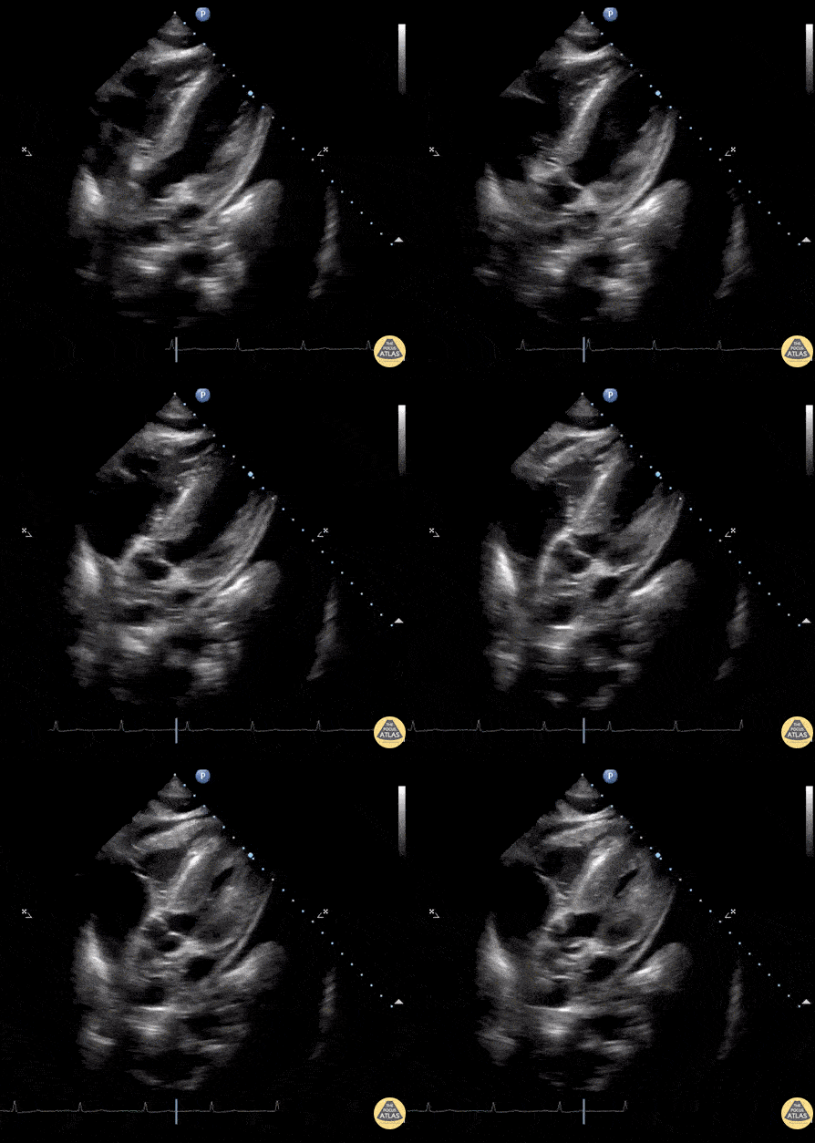
Ascites, Pericardial Effusion & Left Pleural Effusion
Subcostal window of patient with Lupus. An anechoic region is seen within the peritoneum, pericardium, and left lung indicating simultaneous ascites, pericardial effusion, and pleural effusion respectively.
Image courtesy of Robert Jones DO, FACEP @RJonesSonoEM
Director, Emergency Ultrasound; MetroHealth Medical Center; Professor, Case Western Reserve Medical School, Cleveland, OH
View his original post here

Circumferential Pericardial Effusion with Tamponade
Seen here is the subcostal view of a patient with hemodynamic compromise. Note the circumferential area of anechoic fluid, resulting in dynamic right atrial and ventricular collapse as well as global hyperdynamic systolic function. It is important to remember that systolic right atrial collapse may be the earliest echocardiographic sign of tamponade. Diastolic right ventricular collapse indicates decreased cardiac output as a consequence of inappropriate right ventricular filling.
Rupinder Sekhon, MD
Central Michigan University, Emergency Medicine

Cardiac Tamponade secondary to Type A Aortic Dissection
A 72-year-old male presented with syncope and was found to be hypotensive. POCUS revealed a large, circumferential pericardial effusion with tamponade physiology. Additional workup including chest CT revealed POCUS findings to be the consequence of a Type A aortic dissection (with blood extending back into the pericardial sac).
Dr. John Cook, @J_County
Dr. Tim Scheel, @tscheelEMUS

Pre- & Post- Pericardiocentesis
Subcostal view of a pericardial effusion with cardiac tamponade and its resolution with pericardiocentesis.
Renato Melo, Pocus Jedi co-founder, @JediPocus
Emergency Physician at H.C. de Marília/SP Brazil

Pericardial Effusion noted during CPR
POCUS obtained during a cardiac arrest demonstrates both CPR in progress as well as a large pericardial effusion. This patient had been waiting for CT aortogram to further evaluate severe back pain prior to onset of arrest. The pericardial collection appears hyperechoic indicating clotted blood (most likely secondary to a Type A aortic dissection). An attempt at needle aspiration was unsuccessful and the patient died.
Peter Cheng

Attempted Pericardiocentesis
Patient had PEA arrest. View of the heart revealed pericardial effusion. The needle entering the pericardium can been seen in this clip.
Image courtesy of Robert Jones DO, FACEP @RJonesSonoEM
Director, Emergency Ultrasound; MetroHealth Medical Center; Professor, Case Western Reserve Medical School, Cleveland, OH
View his original post here

Confirmation of Needle Placement For Pericardiocentesis
Initial stage of the pericardiocentesis. Agitated saline was injected to confirm needle location prior to withdrawing bloody fluid.
Image courtesy of Robert Jones DO, FACEP @RJonesSonoEM
Director, Emergency Ultrasound; MetroHealth Medical Center; Professor, Case Western Reserve Medical School, Cleveland, OH
View his original post here

Pericardial Cyst
This subcostal view shows cysts located on the pericardium. A benign and rare finding to be aware of while performing an echo.
Image courtesy of Robert Jones DO, FACEP @RJonesSonoEM
Director, Emergency Ultrasound; MetroHealth Medical Center; Professor, Case Western Reserve Medical School, Cleveland, OH
View his original post here

RV Diastolic Collapse in Cardiac Tamponade
A patient with a history coronary artery disease presented to our emergency department complaining of acute weakness. He was febrile, tachycardic and hypotensive.
POCUS was performed and demonstrated a circumferential anechoic rim in multiple views, consistent with a pericardial effusion (subcostal four-chamber view is shown above). Right ventricular (RV) diastolic collapse was visualized and consistent with the diagnosis of pericardial tamponade. Loss of central pulses prompted resuscitative efforts and an emergent pericardiocentesis, which resulted in return of spontaneous circulation.
Andrew Namespetra, MB BCh BAO. @AndrewNamespet1
PGY-1 EM Resident at Central Michigan University
Samantha Wong, DO.
PGY-3 EM Resident at Central Michigan University

Subxiphoid View of Pericardial Effusion
Seen here is a subxiphoid view of a circumferential pericardial effusion. Of note, there is no diastolic RV collapse, therefore no echocardiographic tamponade.
Moudi Hubeishy
@moudihubeishy

Circumferential Pericardial Effusion
Seen here is a circumferential pericardial effusion identified in a patient without hemodynamic evidence of tamponade.
Moudi Hubeishy
@moudihubeishy

Traumatic Tamponade
27-year-old woman presents with gunshot wound to right chest. Patient phonating on arrival but tachycardic in 150s with BP 60/palp. EFAST revealed pericardial effusion with hematoma and near complete RV diastolic collapse, consistent with cardiac tamponade. Decision was made to take patient directly for OR thoracotomy based on EFAST findings. Patient lost pulses for 10 sec intra-operatively prior to receiving pericardial window. ROSC was achieved with removal of pericardial hematoma. LV laceration was identified and repaired. LAD laceration identified for which patient received emergent CABG by CT surgery. Patient was successfully extubated the next day, discharged one week later.
EFAST aids in rapid diagnosis and expedites care in hemodynamically unstable trauma patients. POCUS is a useful tool for determining etiology of undifferentiated shock.
Quinn Fujii DO, William Hamrick DO, Ulysses Garcia DO, Robert Rigby DO
Desert Regional Medical Center, Emergency Medicine

Cardiac Tamponade
Seen here is a parasternal long axis view of a patient with cardiac tamponade.
Francisco Norman

Hemopericardium
POCUS revealed isoechoic fluid within the pericardium indicative of a hemopericardium in a patient suffering from a gunshot wound.
Image courtesy of Robert Jones DO, FACEP @RJonesSonoEM
Director, Emergency Ultrasound; MetroHealth Medical Center; Professor, Case Western Reserve Medical School, Cleveland, OH
View his original post here

Ascitic Fluid on Subcostal View
This subcostal view shows a patient with ascites, a small pericardial effusion, and a left sided pleural effusion. When suspecting a pericardial effusion, always ensure which side of the diaphragm the fluid presents.
Image courtesy of Robert Jones DO, FACEP @RJonesSonoEM
Director, Emergency Ultrasound; MetroHealth Medical Center; Professor, Case Western Reserve Medical School, Cleveland, OH
View his original post here

Intrapericardial Hematoma
Parasternal short axis view of a Post-CABG intrapericardial hematoma.
Image courtesy of Robert Jones DO, FACEP @RJonesSonoEM
Director, Emergency Ultrasound; MetroHealth Medical Center; Professor, Case Western Reserve Medical School, Cleveland, OH
View his original post here

Hemopericardium
POCUS revealed a retained bullet near the left ventricular wall in a patient following a gunshot wound.
Image courtesy of Robert Jones DO, FACEP @RJonesSonoEM
Director, Emergency Ultrasound; MetroHealth Medical Center; Professor, Case Western Reserve Medical School, Cleveland, OH
View his original post here

Pericardial Effusion
Parasternal short axis view of a pericardial effusion. View the thread at the link below for parasternal long axis and associated EKG findings.
Image courtesy of Robert Jones DO, FACEP @RJonesSonoEM
Director, Emergency Ultrasound; MetroHealth Medical Center; Professor, Case Western Reserve Medical School, Cleveland, OH
View his original post here

Uremic Pericarditis
PLAX view in a patient who presented to the ED after missing several scheduled dialysis sessions reveals a small pericardial effusion with a pericardial friction rub on exam.
Image courtesy of Robert Jones DO, FACEP @RJonesSonoEM
Director, Emergency Ultrasound; MetroHealth Medical Center; Professor, Case Western Reserve Medical School, Cleveland, OH
View his original post here

Hemopericardium
A young previously healthy male presented to the ED with a transmediastinal gunshot wound. FAST exam revealed hemopericardium on the subxiphoid window.
Image courtesy of Robert Jones DO, FACEP @RJonesSonoEM
Director, Emergency Ultrasound; MetroHealth Medical Center; Professor, Case Western Reserve Medical School, Cleveland, OH
View his original post here

Fibrinous Pericardial Effusion
A young male presented with a 5-month history of signs and symptoms of heart failure. Apical four chamber view pictured here revealed a large pericardial effusion, most significant for the presence of fibrinous stranding. Fibrinous pericardial effusions are most commonly related to tuberculosis (worldwide) whereas malignancy and inflammatory/infectious processes are alternate possible etiologies. PCR of pericardial fluid went on to confirm Mycobacterium tuberculosis in this patient.
Note: depth of this image was intentionally used to augment the view of the apex and presence of fibrinous strands.
Gordon Johnson, @pdxfutebol
Portland, Oregon

Pericardial Effusion vs Tamponade
Seen here is a subxiphoid view of a moderate sized, free-flowing pericardial effusion. There is >50% collapsible IVC which makes tamponade less likely in the absence of clinical signs of hemodynamic deterioration (i.e. normal blood pressure, mild tachycardia and pulsus paradoxus <10 mmHg).
Plethora of the IVC in pericardial effusion is associated with elevated right heart filling pressures and is more sensitive though less specific for tamponade than right heart chamber collapse or jugular venous distention. Plethora of the IVC also has greater sensitivity than does elevated jugular venous distention on physical examination (97% versus 61%).
Reference: Ronald Himelman, Barbara Kircher, Don Rockey, Nelson Schiller. Inferior vena cava plethora with blunted respiratory response: A sensitive echocardiography sign of cardiac tamponade. JACC. 1988; 12:1470-1477.
Shahad Al Chalaby, MD (PGY3) @shahad_Chalaby
Alameda Health System Internal Medicine Residency Program
Oakland, CA

Constrictive Pericarditis
A young patient with a history of SLE presented to the ED with new onset HFpEF. A subcostal view revealed constrictive pericarditis with septal wall bouncing and a mild pericardial effusion.
Image courtesy of Robert Jones DO, FACEP @RJonesSonoEM
Director, Emergency Ultrasound; MetroHealth Medical Center; Professor, Case Western Reserve Medical School, Cleveland, OH
View his original post here

Purulent Pericarditis
A male in his 20’s presented following failed community acquired pneumonia treatment with fever, chest pain, and soft vitals. Bedside ultrasound revealed purulent pericarditis visible in this subcostal window. CT showed LLL pneumonia with empyema.
Image courtesy of Robert Jones DO, FACEP @RJonesSonoEM
Director, Emergency Ultrasound; MetroHealth Medical Center; Professor, Case Western Reserve Medical School, Cleveland, OH
View his original post here

Cardiac Tamponade with Swinging Heart
31-yo-male presented s/p syncopal episode and ongoing hypotension. Initial evaluation included POCUS that revealed a circumferential pericardial effusion causing RA and RV diastolic collapse as well as a “swinging heart”. Patient was promptly sent for a pericardial window.
Tessa W. Damm, DO
Intensivist, Critical Care Medicine & Neurocritical Care
Wisconsin, USA
@DrDamm
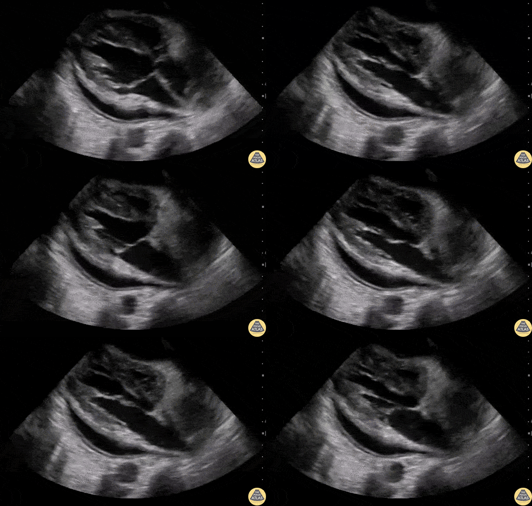
Cardiac Tamponade
60 y/o M with metastatic lymphoma presented with SOB over 3 days. POCUS revealed a pericardial effusion that, along with his vital signs and clinical picture, altogether suggested pericardial tamponade.
Notice the diastolic collapse of the right ventricle (as the mitral valve opens, the right ventricle free wall collapses). Mitral and tricuspid inflow velocities were also used as a surrogate for pulsus paradoxus.
Dr. Stephen Alerhand - Mt Sinai Hospital, NYC

Acute Pericarditis
A young male presented with 2-day history of chest pain. His ECG revealed diffuse ST segment elevation. He had an elevated but serially stable troponin level. POCUS excluded focal wall motion abnormality though was notable for the presence of a small pericardial effusion and a thickened, hyperechoic pericardium; consistent with his diagnosis of acute pericarditis.
Renato Melo, Emergency Physician
@Renato_Melo_

Cardiac Tamponade in PEA
A 83-year-old woman comes to emergency department In cardiac arrest after an episode of severe chest pain. During pulse check we identified PEA (Pulseless Electrical Activity) and performed a CASA Exam. The subcostal view demonstrates a hyperechoic structure in the pericardial space concerning for clot. There is also evidence of cardiac tamponade.
Image courtesy of Josiane Almeida

Pericardial Effusion in Parasternal Short-Axis
Here is an example of relatively preserved myocardial function in the setting of a moderate-sized, circumferential pericardial effusion, as seen from the parasternal short-axis view.
Edgar Miranda, MD
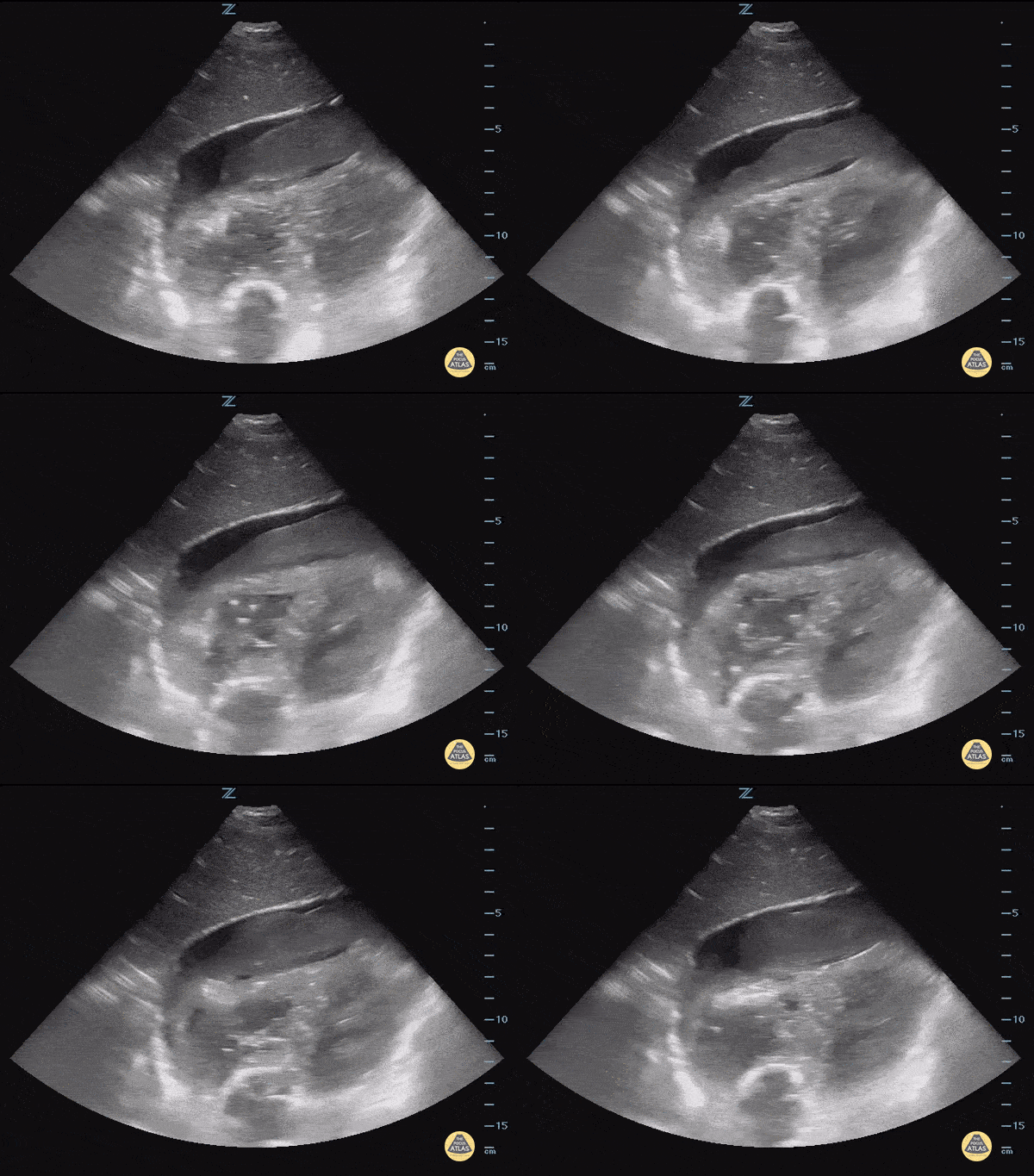
Pericardial Effusion with Hematoma
This is a subcostal view of a patient presenting after a motor vehicle collision. The image demonstrates a pericardial effusion containing a large hypoechoic mobile structure concerning for hematoma.
Image courtesy of IUEM Ultrasound
Original Twitter post can be found here.

Pericardial Effusion and Rapid Atrial Fibrillation
50 y/o male presented with 1 week of feeling generally unwell, tired and short of breath. Rapid atrial fibrillation was present in the department. POCUS performed demonstrating large pericardial effusion (note some evidence of diastolic collapse as well as partially complex-appearing pericardial fluid collection).
Nishant Cherian
Emergency Medicine Registrar

Pericardiocentesis
Dynamic needle guidance in a parasternal view used to perform an emergent pericardiocentesis in a hemodynamically unstable patient.
Ahad Al Saud, Emergency Medicine Physician @Ahad_AlSaud
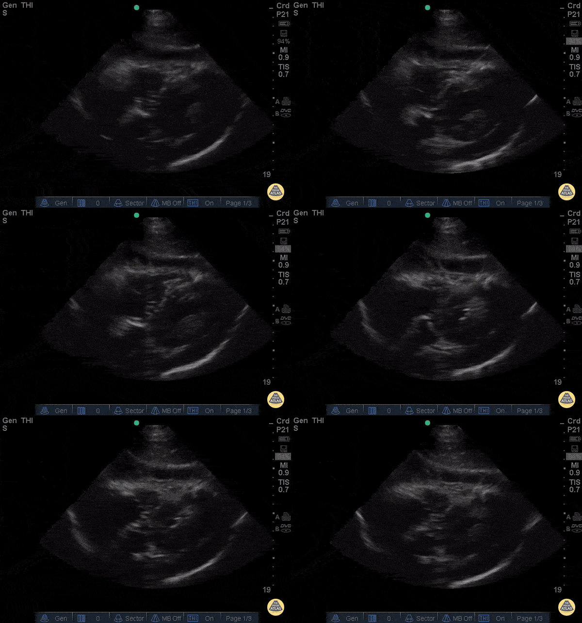
Exudative Effusion
A 67y/o M PMH ESRD presented to the ED with recurrent sharp chest pain, cough, and fever that were reported during routine dialysis. He had been recently hospitalized for chest pain one week prior and diagnosed with pericardial effusion. Upon repeat presentation to the ED, the patient’s vital signs and examination were unremarkable. ECG showed diffuse PR depressions and ST elevations. PoCUS (cardiac subcostal view shown) demonstrated a circumferential, anechoic pericardial fluid collection with septations; there was no evidence of tamponade.
The patient was started on indomethacin and colchicine and admitted to the CCU. A few days later, cardiothoracic surgery performed a pericardial window, drained 350 ml of serosanguinous fluid, and lysed multiple adhesions. Biopsied tissue revealed a thickened, inflamed pericardium with abscess and granulation tissue formation. Pericardial fluid testing was negative for bacteria, fungi, acid-fast bacilli, and malignancy.
Emergency physicians can detect pericardial effusion with a sensitivity of 96% (95% confidence interval [CI] 90.4% to 98.9%), specificity of 98% (95% CI 95.8% to 99.1%).[i] High risk features that warrant hospital admission include temperature > 38°C, subacute course, large effusion (echo-free space > 20 mm), tamponade, and lack of response to aspirin or non-steroidal anti-inflammatory drug therapy; however, the etiology often remains a mystery. [ii] The presence of a loculated effusion on echocardiography may suggest the eventual need for surgical intervention with pericardiectomy or pericardial window.[iii]
Dr. Ian Desouza - Kings County Emergency Medicine
[i] Mandavia DP, Hoffner RJ, Mahaney K, Henderson SO. Bedside echocardiography by emergency physicians. Ann Emerg Med 2001;38:377-82.
[ii] Imazio M, Cecchi E, Demichelis B, et al. Indicators of poor prognosis of acute pericarditis. Circulation 2007;115:2739-44.
[iii] Imazio M, Spodick DH, Brucato A, Trinchero R, Adler Y. Controversial issues in the management of pericardial diseases. Circulation 2010;121:916-28.
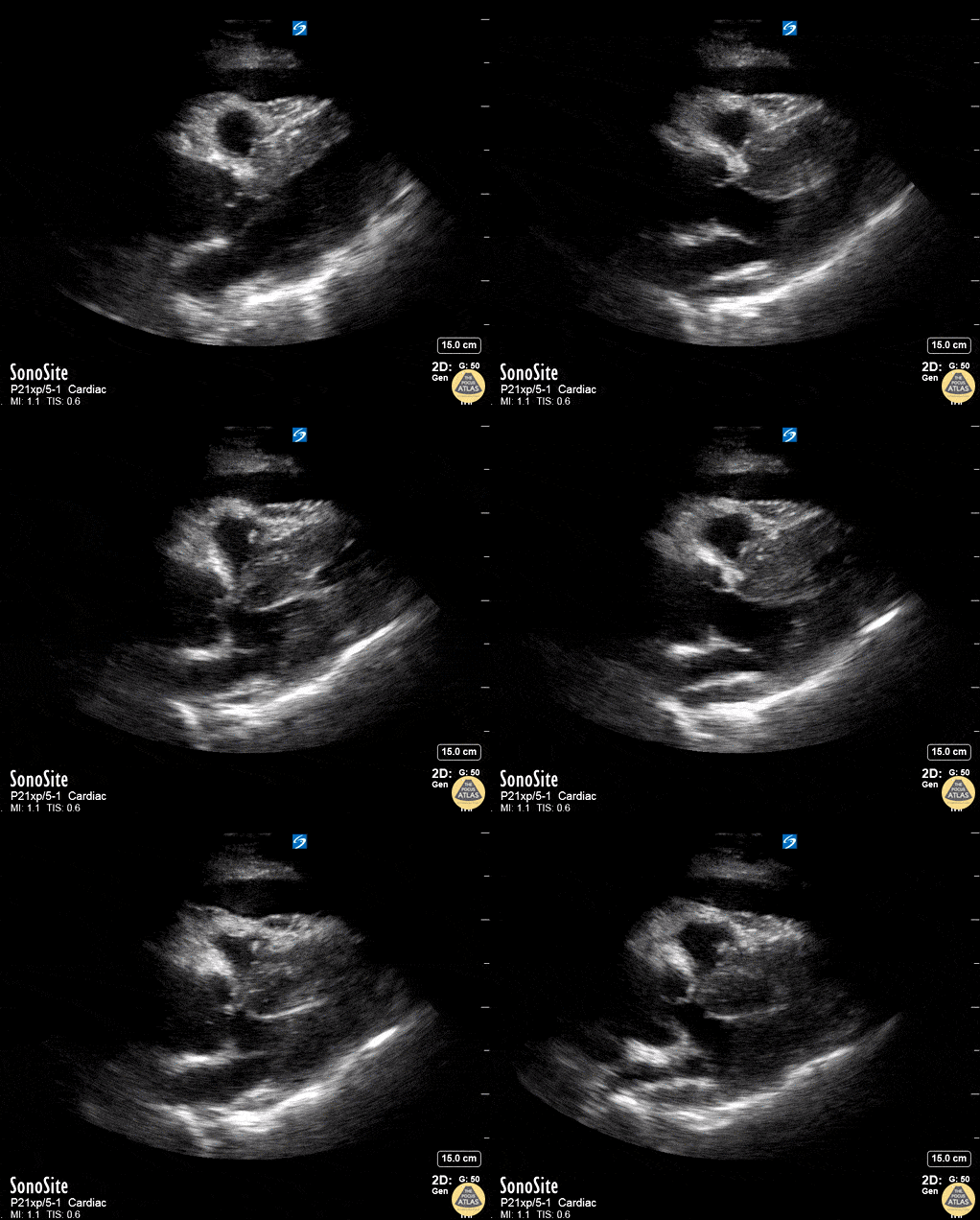
Aortic Dissection with Tamponade - Parasternal Long
50 year old mandarin speaking man complains of vague central chest pain. You pursue a routine cardiac workup which is fairly normal. Upon discharging him, the nurse tells you his systolic in now in the 80's.
You perform a RUSH exam - echo demonstrates a widened aortic outflow tract (well exceeding the rule of thirds) suggesting a Type A aortic dissection. You also see a pericardial effusion with right ventricular diastolic collapse. This is "the man jumping on the trampoline" or dimpling of the right ventricular free wall during diastole while it should be filling - clenching the diagnosis of tamponade.
You better hope your CT surgeon is in house. Scroll to see the rest of the case for further images.
Dr. Matthew Riscinti - Kings County Emergency Medicine, Dr. Benjamin Clearly - NYU Langone Emergency Medicine

Aortic Dissection with Tamponade - Apical
50 year old mandarin speaking man complains of vague central chest pain. You pursue a routine cardiac workup which is fairly normal. Upon discharging him, the nurse tells you his systolic in now in the 80's.
Your RUSH exam and echo on this apical four demonstrate a pericardial tamponade with both:
1. Right ventricular DIASTOLIC collapse - dimpling of the right ventricular free wall during diastole while it should be filling
2. Right Atrial SYSTOLIC collapse - (but in atrial diastole) while the atrium should be filling
Scroll to see the rest of the case for further images.
Dr. Matthew Riscinti - Kings County Emergency Medicine, Dr. Benjamin Clearly - NYU Langone Emergency Medicine
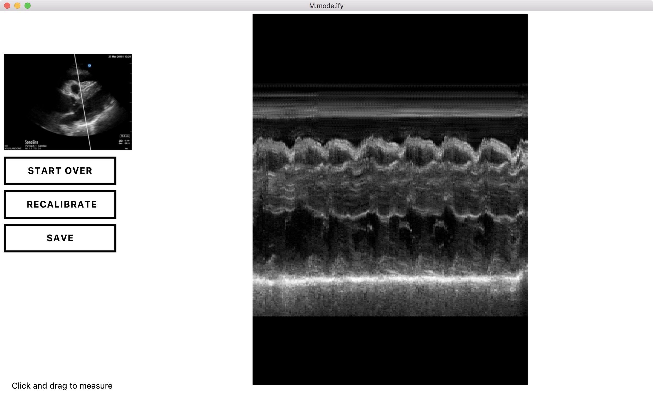
Aortic Dissection with Tamponade - M-mode
Your RUSH exam's echo, parasternal long, shows tamponade. M-mode can be used to confirm by placing the M-mode line through the RV and the mitral valve to better visualize the RV diastolic collapse. The top line of the image shows the RV free wall collapsing during diastole when it should be filling. You know this is during diastole because the mitral valve is opening at this time, seen toward the bottom of the M-mode image.
This image was created retrospectively using M.Mode.ify by Dr. Ben Smith.
Dr. Matthew Riscinti - Kings County Emergency Medicine, Dr. Benjamin Clearly - NYU Langone Emergency Medicine
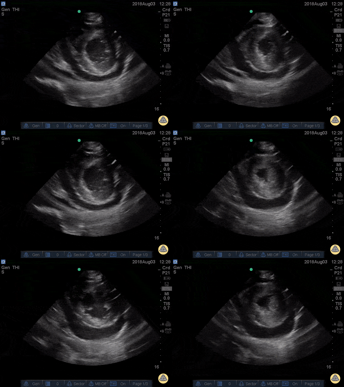
Cardiac Tamponade Parasternal Short
41 yo M with history of stage 4 lung cancer presents with AMS and dyspnea, normotensive and tachycardic to 140s.
Parasternal long view showed moderate pericardial effusion with RV collapse. With M mode we are able to see the RV wall collapse (top line) corresponds with the mitral valve opening i.e. it occurs during diastole. Even though the patient was normotensive he was taken to the OR for a pericardial window within the hour given this evidence of echocardiographic tamponade.
Nathan Kabariti MS4, Dr. Charles Murchison, Dr. John Riggins, Dr. Donald Doukas - Kings County Emergency Medicine
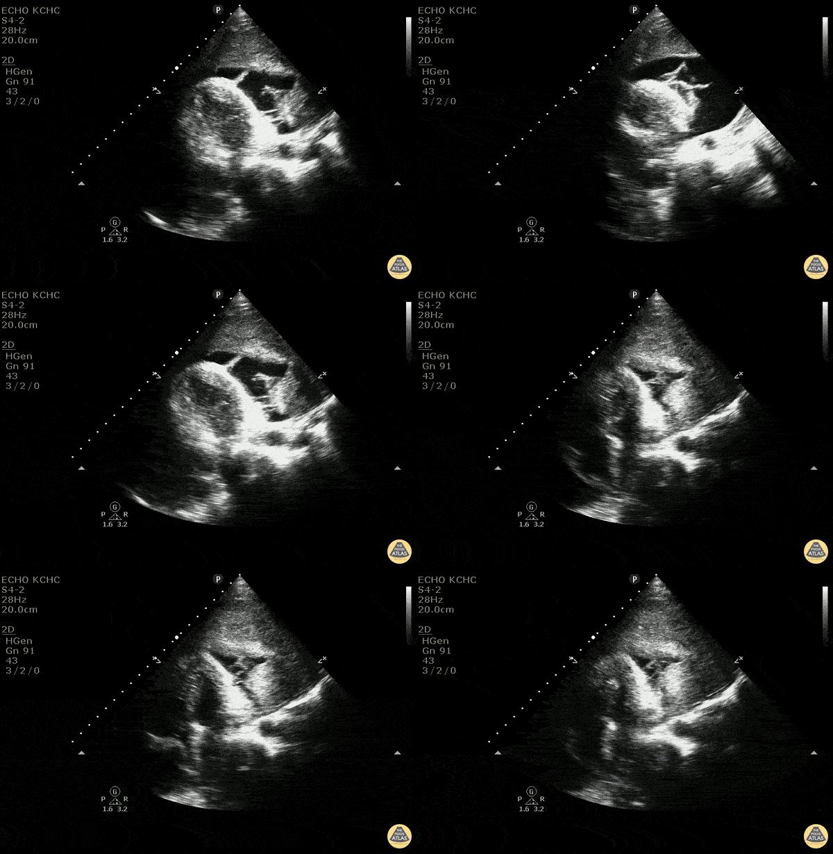
Malignant Pericardial Effusion
40 y/o F with 2-3 months of weight loss and new diagnosis of mediastinal mass. Sent from clinic for SOB. Patient initially tachycardic, tachypnic. EKG shows electrical alternans.
US shows large hypoechoic area with mobile hyperechoic lines in an effusion concerning for fibrinous exudate/growth. There is no sign of end diastolic right ventricular collapse. Findings are consistent with a pericardial effusion with fibrin deposits without evidence of tamponande.
For POCUS - the easiest assessment of tamponade is to look at end diastolic right atrial and ventricular collapse. Since the right side of the heart typically is at lower pressures, it will be the first to collapse from the pressure in the pericardium. It is easily measured with m-mode across the wall of the ventricle or atria. Bedside US is an amazing tool and can quickly and effectively show the hemodynamic status in pericardial effusion, so that the correct management can be done.
Dr. Carolina Camacho, Taylor Surles, Michael Greisinger, and Scott Kendall - Kings County Emergency Medicine
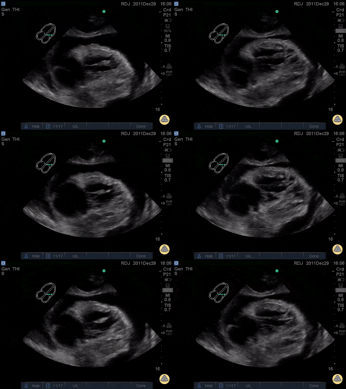
Chronic Pericardial Effusion
WCUME17 submission for "Creative Caption"
"When the turtle's head appears you know you're in trouble!"
Patient with chronic pericardial effusion and... a heart that looks like a snapping turtle?
Dr. Robert Jarman - UK
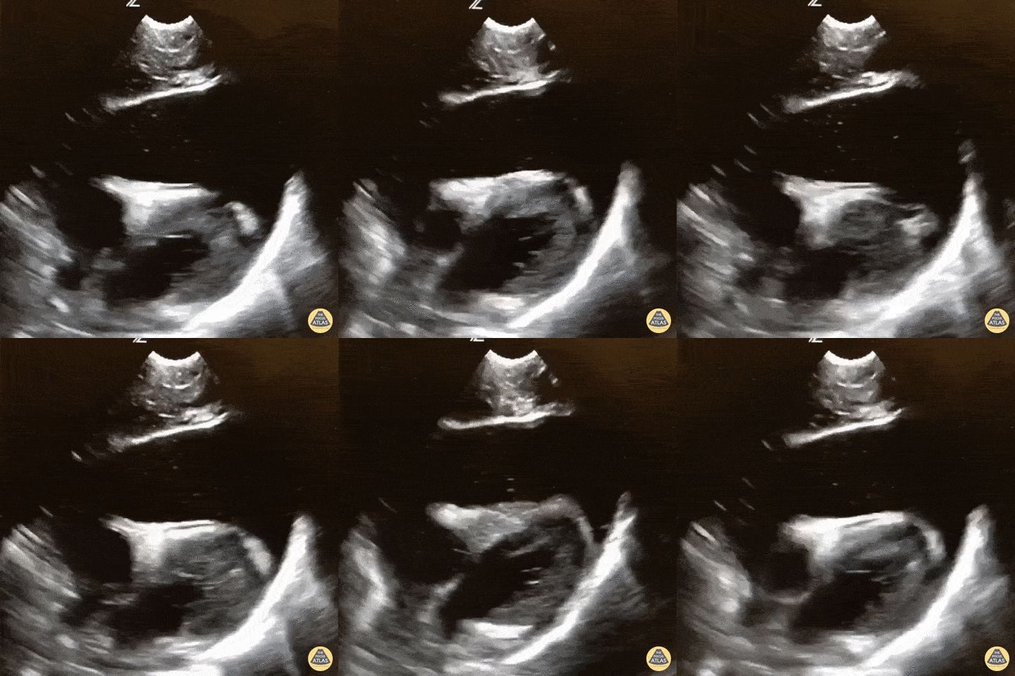
Acute Traumatic Cardiac Tamponade
WCUME 2017 Submission for "Best POCUS"
Massive hemopericardium with coagulating blood and tamponade in a pediatric trauma patient. The patient went straight to the OR based on this image!
Dr. Sarah Medeiros - Sacramento, CA
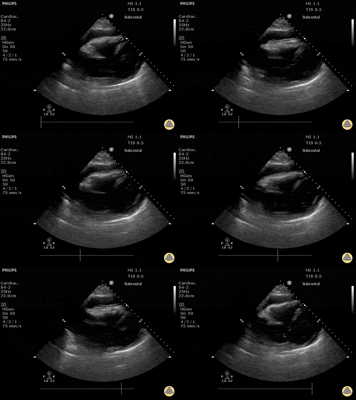
Cardiac Tamponade - Subxiphoid View
This is a subxiphoid view demonstrating a moderate pericardial effusion and diastolic collapse of the right ventricle concerning for cardiac tamponade.
Justin Bowra MBBS, FACEM, CCPU Emergency Physician, RNSH et al.
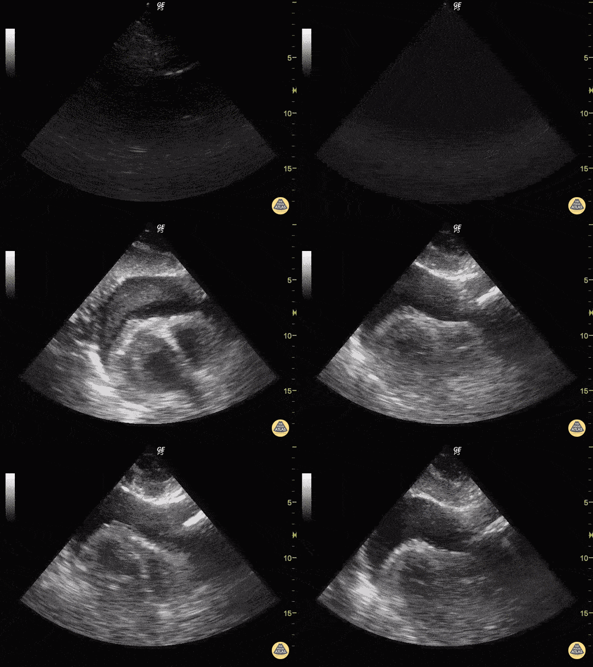
Cardiac Tamponade in Stabbed Patient
Hematoma and relatively hypoechoic blood can be seen around the heart.
Justin Bowra MBBS, FACEM, CCPU Emergency Physician, RNSH et al.
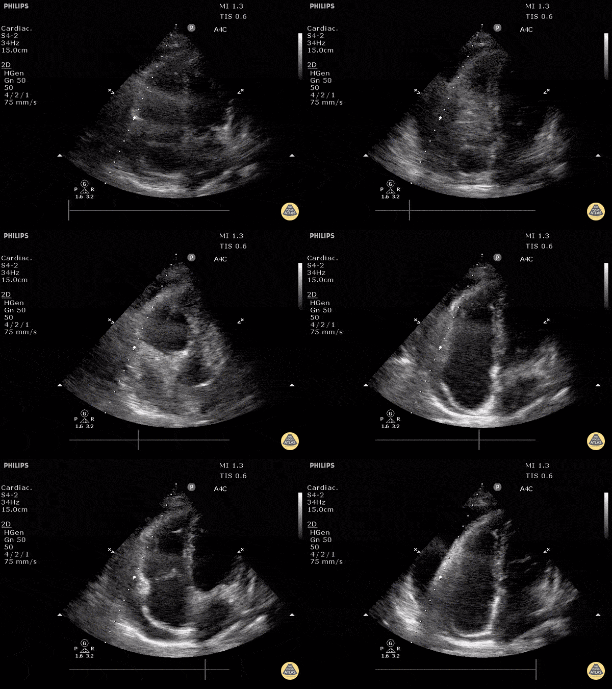
Cardiac Tamponade in Apical View
Apical view demonstrating rocking heart with diastolic collapse of RV free wall.
Justin Bowra MBBS, FACEM, CCPU Emergency Physician, RNSH et al. (Dr. Orr)
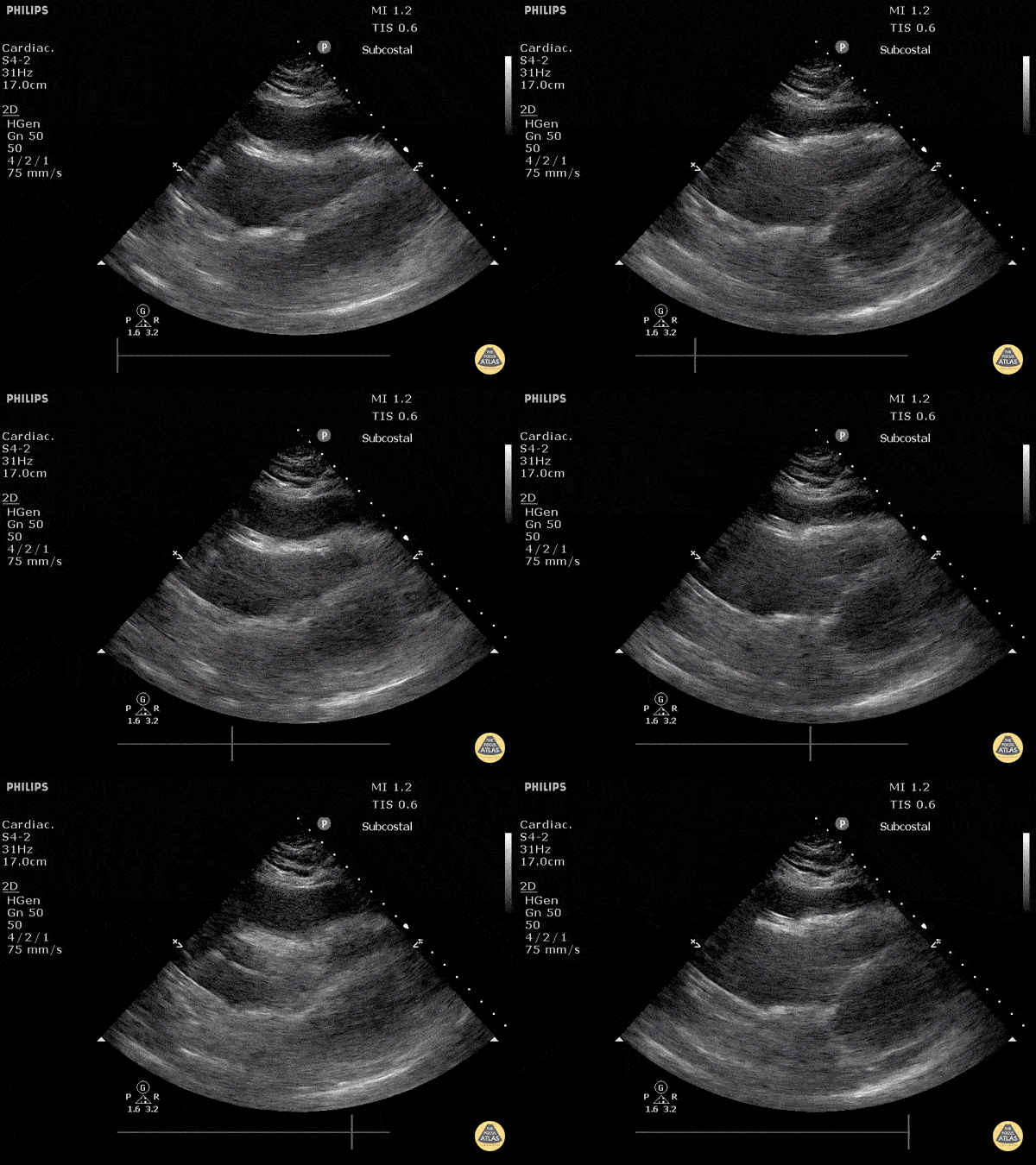
Cardiac Tamponade
Subcostal view demonstrating signs of cardiac tamponade including right ventricular collapse during diastole.
Justin Bowra MBBS, FACEM, CCPU Emergency Physician, RNSH et al. (Dr. Orr)
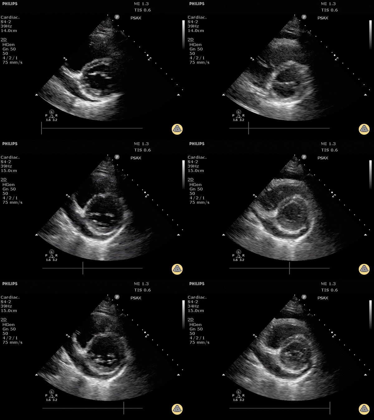
Cardiac Tamponade (Parasternal Short-Axis View)
Parasternal short axis view demonstrating evidence of diastolic RV free wall collapse.
Justin Bowra MBBS, FACEM, CCPU Emergency Physician, RNSH et al. (Dr. Orr)
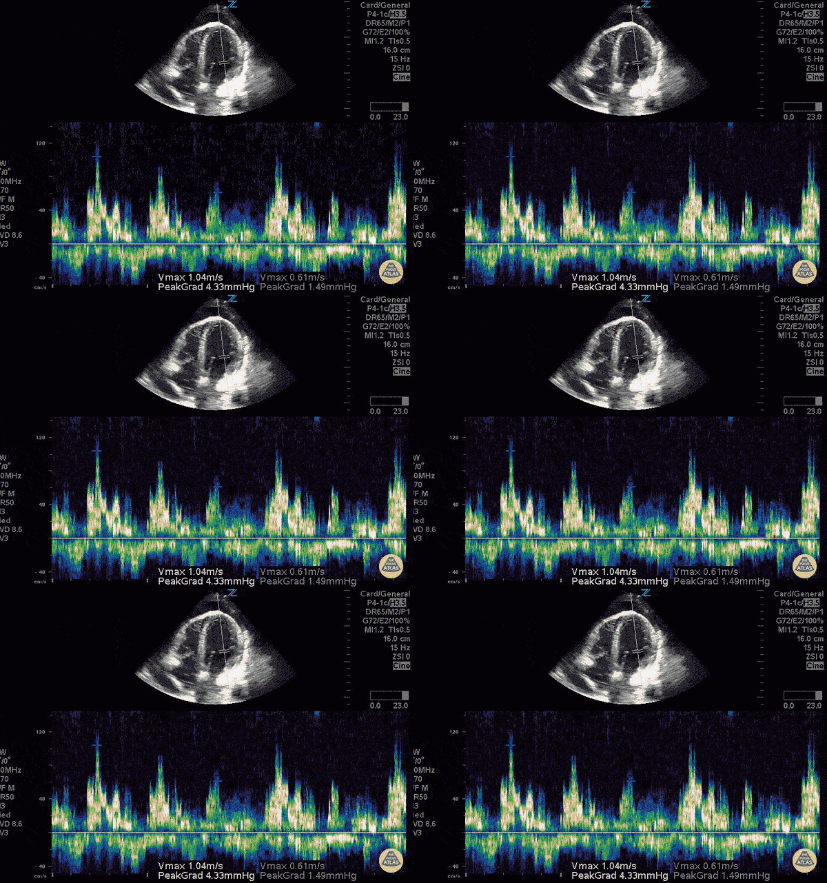
Cardiac Tamponade with Doppler
Mitral inflow velocity respiratory variation > 25% consistent with cardiac tamponade physiology.
Sukh Singh, MD
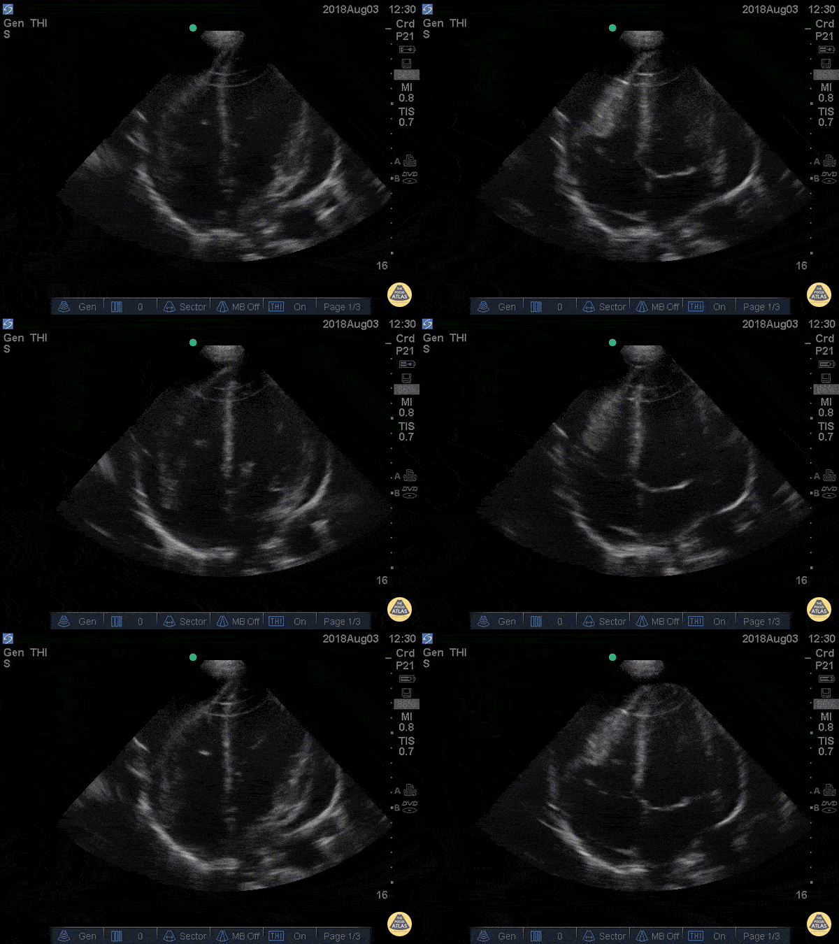
Cardiac Tamponade Apical Four
41 yo M with history of stage 4 lung cancer presents with AMS and dyspnea, normotensive and tachycardic to 140s.
Parasternal long view showed moderate pericardial effusion with RV collapse. With M mode we are able to see the RV wall collapse (top line) corresponds with the mitral valve opening i.e. it occurs during diastole. Even though the patient was normotensive he was taken to the OR for a pericardial window within the hour given this evidence of echocardiographic tamponade.
Nathan Kabariti MS4, Dr. Charles Murchison, Dr. John Riggins, Dr. Donald Doukas - Kings County Emergency Medicine
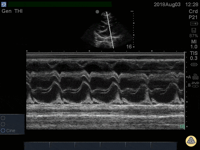
Cardiac Tamponade RV Diastolic Collapse M-mode
41 yo M with history of stage 4 lung cancer presents with AMS and dyspnea, normotensive and tachycardic to 140s.
Parasternal long view showed moderate pericardial effusion with RV collapse. With M mode we are able to see the RV wall collapse (top line) corresponds with the mitral valve opening i.e. it occurs during diastole. Even though the patient was normotensive he was taken to the OR for a pericardial window within the hour given this evidence of echocardiographic tamponade.
Nathan Kabariti MS4, Dr. Charles Murchison, Dr. John Riggins, Dr. Donald Doukas - Kings County Emergency Medicine
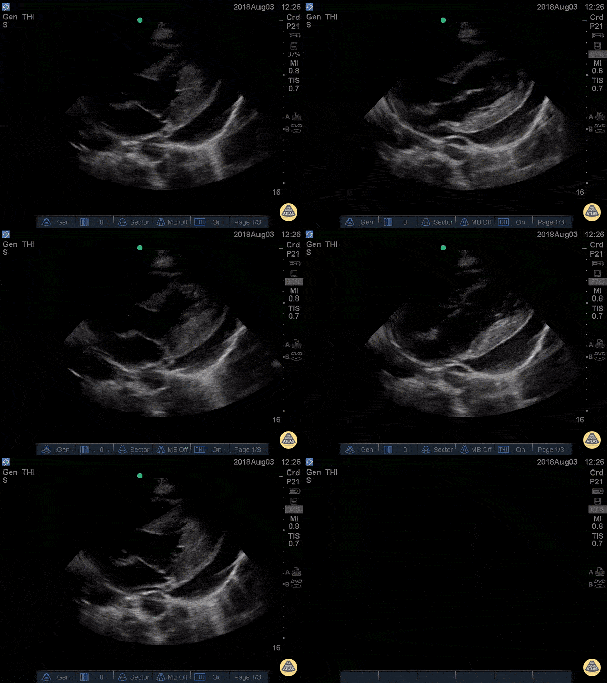
Cardiac Tamponade Parasternal Long
41 yo M with history of stage 4 lung cancer presents with AMS and dyspnea, normotensive and tachycardic to 140s.
Parasternal long view showed moderate pericardial effusion with RV collapse. With M mode we are able to see the RV wall collapse (top line) corresponds with the mitral valve opening i.e. it occurs during diastole. Even though the patient was normotensive he was taken to the OR for a pericardial window within the hour given this evidence of echocardiographic tamponade.
Nathan Kabariti MS4, Dr. Charles Murchison, Dr. John Riggins, Dr. Donald Doukas - Kings County Emergency Medicine
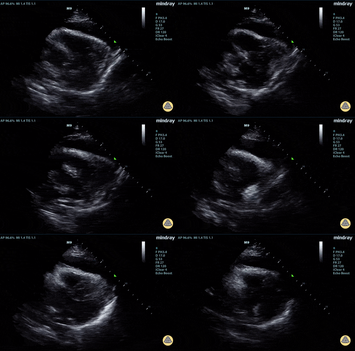
Parasternal Long Type A Aortic Dissection with Tamponade
Elderly fellow who had a headache while bike riding, with some leg weakness. No chest or back pain. Stable for hours then came to hospital, suddenly hypotension and drowsy in ER
POCUS RUSH Exam performed lead to rapid diagnosis of Aortic Dissection with tamponade.
Right ventricular diastolic collapse can be seen.
Claire Heslop - Pediatric Emergency Medicine - University of Toronto Hospital for Sick Children


![Pericardial Effusion Vs Fat Pad [1/2]](https://images.squarespace-cdn.com/content/v1/58118909e3df282037abfad7/1706570222652-Z6J3FZ0G4CX2KG8QEL57/image-asset.gif)
![Pericardial Effusion Vs Fat Pad [2/2]](https://images.squarespace-cdn.com/content/v1/58118909e3df282037abfad7/1706570376077-9Q7LONXIFC3PJAQY5WZJ/image-asset.gif)


