
Right Ventricular Dysfunction
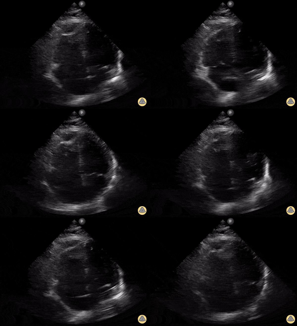
McConnell's Sign Due to NSTEMI
An elderly patient presented with shortness of breath and generalized weakness found to have an NSTEMI and high degree AV block. Bedside focused cardiac ultrasound with 4 chamber apical view revealed: McConnell’s Sign.
Chest CTA neg for PE. Patient underwent cardiac catherization with culprit lesion being mid RCA.
Not all McConnell’s sign is related to acute PE. In this case RV infarct secondary to NSTEMI with mid RCA occlusion s/p DES PCI with complete resolution of AV block.
Dat Lu MD, Internal Medicine Clerkship Site Director Kaiser Roseville

McConnell's Sign on PLAX View
RV dilation violating the rule of thirds. McConnell’s sign is also demonstrated in this view with RV apical hyperkinesis and lateral wall hypo/akinesis. Patient was found to have a submassive pulmonary embolism.
Moudi Hubeishy

Clot in Transit
An 50-year-old presented with shortness of breath and suffered witnessed PEA arrest subsequent to having obtained this POCUS image (subcostal view) notable for a hyperechoic, mobile structure within the RA). ACLS was administered in addition to TPA and ROSC was achieved. Subsequent formal ultrasound, following thrombolytic administration, revealed absence of thrombus.
Ryan Shelby PGY-3 EM Resident; @RyanShelby18
Central Michigan University

Septal Flattening in Saddle Pulmonary Embolism
PSSX View. Septal Flattening is being demonstrated (D-sign) at the level of the papillary muscles and chordae tendinae visible in both the left ventricle and right ventricle. The right ventricle is demonstrating significant dilation in the setting of increased pressures secondary to a saddle embolus.
Moudi Hubeishy
@moudihubeishy

McConnell's Sign
A female in her mid-30’s presented to the ED with chest pain and shortness of breath. POCUS at the apical window revealed right ventricular free wall akinesis with sparing of the apex, indicative of McConnell’s sign of a pulmonary embolism. A clot can also be seen within the right atrium.
Image courtesy of Robert Jones DO, FACEP @RJonesSonoEM
Director, Emergency Ultrasound; MetroHealth Medical Center; Professor, Case Western Reserve Medical School, Cleveland, OH
View his original post here

Obstructive Shock & RUSH
Shown here is an image acquired while performing a RUSH exam on a 40-year-old female who presented in shock. HPI notable for SOB and worsening abdominal distention x5 days; PMH of decompensated etoh cirrhosis. Vitals included BP 80/40; HR 110; RR 30; O2 sat 90% RA.
Seen here is the image obtained while performing subcostal sweep from base to apex. It is most notable for a dilated, hypokinetic RV with paradoxical septal motion and an underfilled, hyperdynamic LV. There is also intraperitoneal free fluid appreciated. Subsequent CT chest confirmed presence of large, bilateral pulmonary emboli as etiology of patient’s shock.
David Carroll

RV Strain
A 39-year-old female with known metastatic lung cancer complicated by multiple pulmonary emboli on apixaban presented with worsening dyspnea and productive cough over one week duration. POCUS identified a large circumferential pericardial effusion in multiple cardiac views in addition to signs of RV strain as demonstrated by flattening of her interventricular septum and ventricular interdependence (shown here on parasternal short axis view). The findings of RV strain are likely chronic and secondary to chronic thromboembolic disease complicated by pulmonary hypertension.
Shahad Al Chalaby, MD. PGY3 Internal Medicine.Highland Hospital. Alameda Health System Residency Program. Oakland, CA, USA. @shahad_Chalaby
Adam Mortimer, DO. PGY1 Internal Medicine. Highland Hospital. Alameda Health System Residency Program. Oakland, CA USA.

D-Shaped Septum
A 56-year-old female with history of metastatic breast cancer presented with acute shortness of breath. Physical examination was notable for hypotension, sinus tachycardia, raised JVP, and mild respiratory distress. There was no evidence of lower extremity edema and chest xray was unremarkable.
POCUS showed no B lines on lung views. Cardiac views (seen here) were notable for a hypokinetic and dilated RV with flattening of the interventricular septum creating a D shaped LV during systole. These findings are consistent with high right ventricle and pulmonary artery pressures in the absence of pulmonary artery stenosis. Specifically, a D-shaped LV during diastole reflects RV volume overload; a D-shaped LV during systole reflects RV pressure overload. RV pressure overload can be seen in massive and submassive pulmonary embolism (PE) and this patient was diagnosed with a submassive PE; findings confirmed by CT angiography. In this instance, POCUS enabled prompt diagnosis and early initiation of therapeutic anticoagulation.
Shahad Al Chalaby, MD. Highland Hospital. Alameda Health System Internal Medicine Residency Program. California, USA. shahad_Chalaby
Katherine Farley, OMS4. Touro University. California, USA.

Clot in Transit
38 yo previously-healthy male 8 days s/p ligamentous repair of the knee, presented with dyspnea and chest pain. Physical exam on room air notable for tachypnea (RR 35), hypoxia (O2 sat 89%), and lungs that were clear to auscultation. POCUS revealed intracardiac thrombus with D-sign and McConnell's Sign. Pt received full-dose anticoagulation prior to CTA-PE confirming the suspected diagnosis of PE. He subsequently underwent successful thrombectomy.
Stacey Frisch, MD
Chief Resident, Emergency Medicine
Kings County Hospital/SUNY Downstate
@emergenStacey
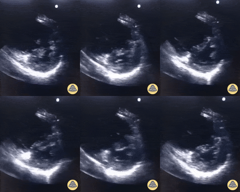
The D-Sign in Acute PE
A 70 year old female patient presented with complaints of acute dyspnea. Oxygen saturation was 86% on room-air improved with O2-supplementation. She had poor hemodynamics with SBP ~ 100 mmHg. POCUS showed obvious right heart strain with a D-sign and thrombus intermittently visualized in the right ventricle. Correct therapy (thrombolytics) were given almost straight away after presentation in the ED because POCUS was available. Once stabilized, the patient was taken for a CTA chest which demonstrated a saddle embolism.
Dr. Van Roosmalen

McConnell's Sign
An elderly female presented with complete AV heart block. Apical 4-chamber view was performed and was notable for regional RV dysfunction (aka McConnell’s Sign). Though this finding is commonly associated with acute PE, our patient was found to have right coronary artery occlusion as the etiology of all findings.
Renato Melo, Emergency Physician at HC de Marília-SP, Brazil. PocusJedi co-founder.
@Renato_Melo_

Measure TAPSE for RV function in PE
This apical 4-chamber view on M-mode was obtained from a patient with acute pulmonary embolism with right ventricular strain.
We measured tricuspid annular plane systolic excursion (TAPSE) as a validated parameter of global RV function. Specifically, TAPSE describes apex-to-base shortening and correlates closely with right ventricular ejection fraction.
TAPSE is measured on echo by placing a cursor at the tricuspid lateral annulus and measuring the distance to systolic annular RV excursion. Any distance of <17mm is suggestive of RV strain. Our patient’s TAPSE was 11.5mm.
Though there are multiple strategies to assess for right heart strain, TAPSE has the highest interrater reliability and best predicts 30-day mortality.
DaMarcus Ingram (MS4), Drexel University College of Medicine
Arthur Strzepka (MS4, Nova Southeastern University College of Osteopathic Medicine
Dr. Max Cooper, Director of Emergency Ultrasound at Crozer Chester Medical Center

RV Thrombus & McConnell's Sign
60 year-old smoker presents with dyspnea and chest pain. Apical 4 chamber view is notable for a thrombus within the RV and associated evidence of RV strain including increased RV size and impaired systolic function with sparing of the apex (known as McConnell’s sign).
Renato Tambelli, @R_Tambelli
Emergency Physician Hospital das Clínicas de Marília, Sao Paulo/Brazil

Post Traumatic Arrest Echo with RA/RV Thrombus
This image is from a patient presenting after a high speed MVC. The patient had a past medical history of atrial fibrillation and was anti-coagulated. On arrival to ED, the patient was agitated, clammy, and noting shortness of breath. The initial eFAST was negative. The patient was intubated with propofol for CT imaging. 15 min post-intubation the patient became hemodynamically unstable and bradycardic followed by cardiac arrest. An apical four chamber view was obtained during pulse check showing dense clot in RV/RA with minimal cardiac activity.
Nishant Cherian
Emergency Medicine Registrar

Right Ventricular Enlargement in Chronic COPD
This clip demonstrates severe right ventricular enlargement seen on a parasternal long axis view. The patient had developed cor pulmonale from longstanding COPD.
Pooja Belligund

Hyperechoic lesion with enlarged RV from PE
[SonoClipShare Submission]: PLA demonstrating a small LV, an enlarged RV, and a hyperechoic lesion moving back and forth (later views confirming moving between RA and RV). 1st of 4 views to be submitted on the same case. This was an unfortunate 40ish y/o 6 months s/p CVA 2/2 ruptured aneurysm and IDDM presenting with 4 days of "feeling weak". Found to have a HR of 120 to 130, a BP around 90-100/60-70 and pulse ox of 90 - 92%. POCUS lungs were normal. Started on heparin, CTA confirmed PE and went to IR for catheter-directed tPA lysis with normalization of BP, HR and good clinical outcome.
Image courtesy of John Hipskind MD
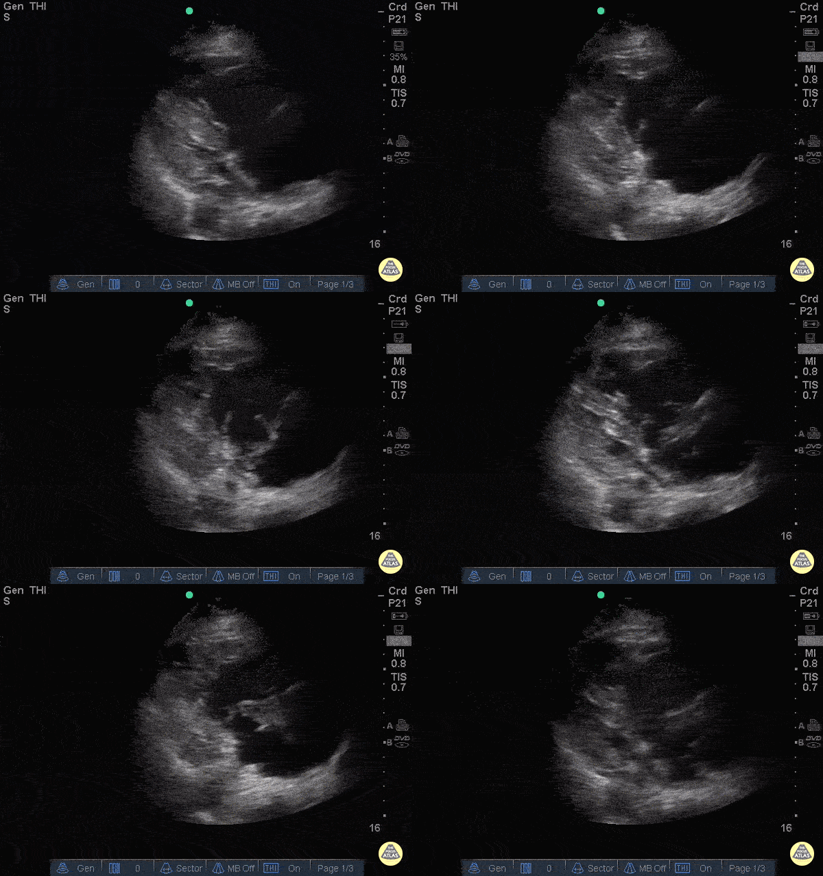
Right Heart Thrombus pre TPA
88 y/o F presenting with chest pain and syncope. Shortly after arrival patient went into brief period of cardiac arrest with ROSC. POCUS shows massive thrombus floating back and forth across the tricuspid valve with a dilated right ventricle, D-sign, and global right heart hypokinesis. Patient was given TPA bolus. Approximately 1 hour after pushing TPA, repeat POCUS with resolution of thrombus on RV, but patient continued RV hypokinesis. Patient vitals stabilized enough for a CTA which showed left main pulmonary artery extending into both left upper lobe and left lower lobe branches with evidence for right heart strain.
Dr. Roderick Alfonso, Maan Al Dubayan, Andrew Sweeny - Kings County Emergency Medicine
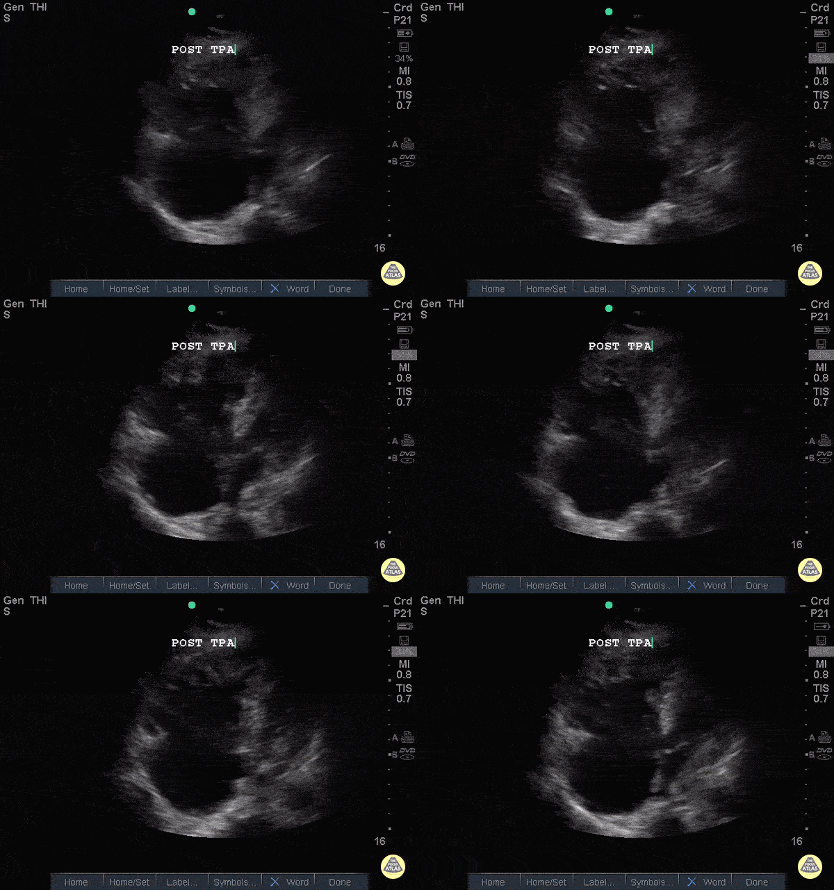
Right Heart Thrombus post TPA
88 y/o F presenting with chest pain and syncope. Shortly after arrival patient went into brief period of cardiac arrest with ROSC. POCUS shows massive thrombus floating back and forth across the tricuspid valve with a dilated right ventricle, D-sign, and global right heart hypokinesis. Patient was given TPA bolus. Approximately 1 hour after pushing TPA, repeat POCUS with resolution of thrombus on RV, but patient continued RV hypokinesis. Patient vitals stabilized enough for a CTA which showed left main pulmonary artery extending into both left upper lobe and left lower lobe branches with evidence for right heart strain.
Dr. Roderick Alfonso, Maan Al Dubayan, Andrew Sweeny - Kings County Emergency Medicine
McConnell's Sign - Pulmonary Embolism
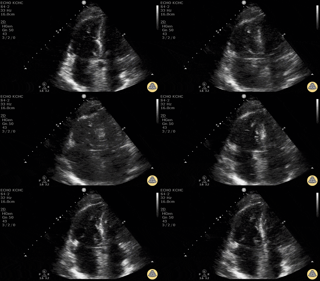
Elderly patient with chest pain, sob, and HR in 120’s. During ER stay became hypotensive with systolic in 80’s. POCUS demonstrated RV enlargement and McConnell’s Sign - systolic akinesia of the RV free wall with preserved functioning of the apex. This is concerning for acute PE. Patient became progressively hypotensive and TPA was pushed.
Dr. Kelly Maurelus, Matthew Riscinti - Kings County Emergency Medicine
Right-Sided Heart Failure (A4C)
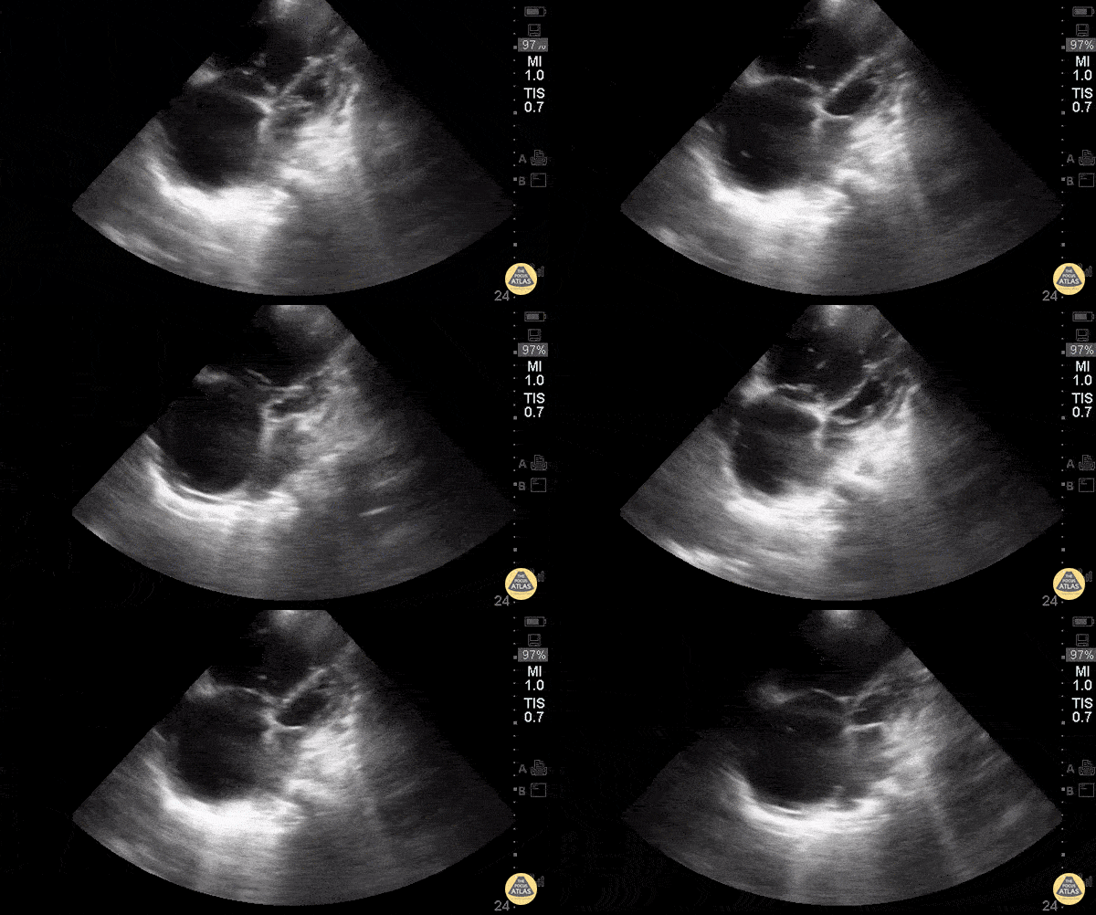
Young woman with a history of Idiopathic Pulmonary Arterial Hypertension w/ resultant R heart failure who came in short of breath.
Greg Powell, MD
Right-Sided Heart Failure (PSAX)
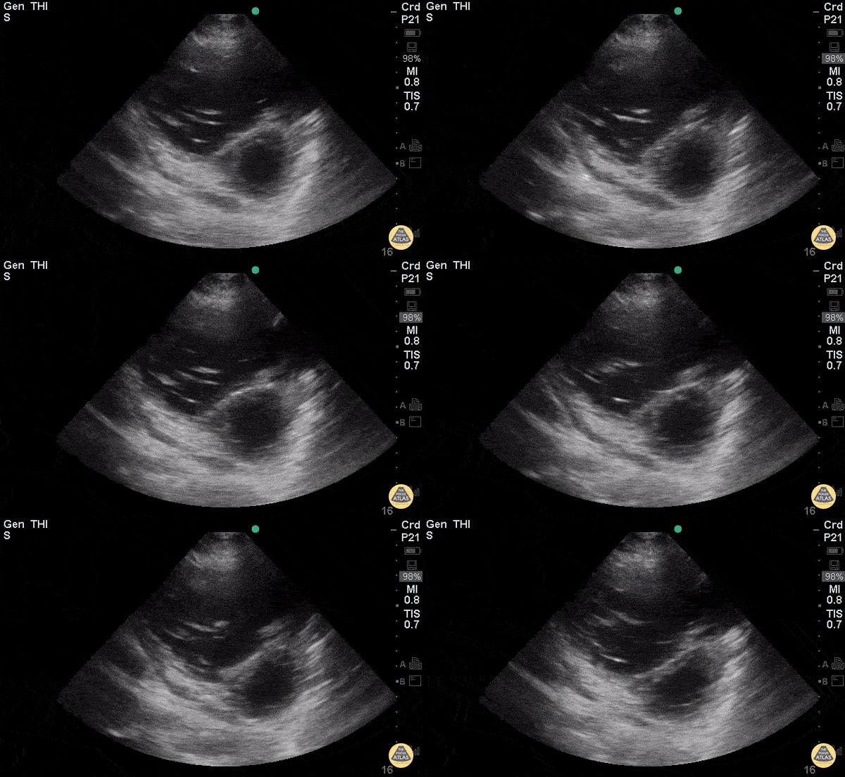
Young woman with a history of Idiopathic Pulmonary Arterial Hypertension with resultant R heart failure who came in short of breath.
Greg Powell, MD
D-Sign in acute PE - Pre TPA
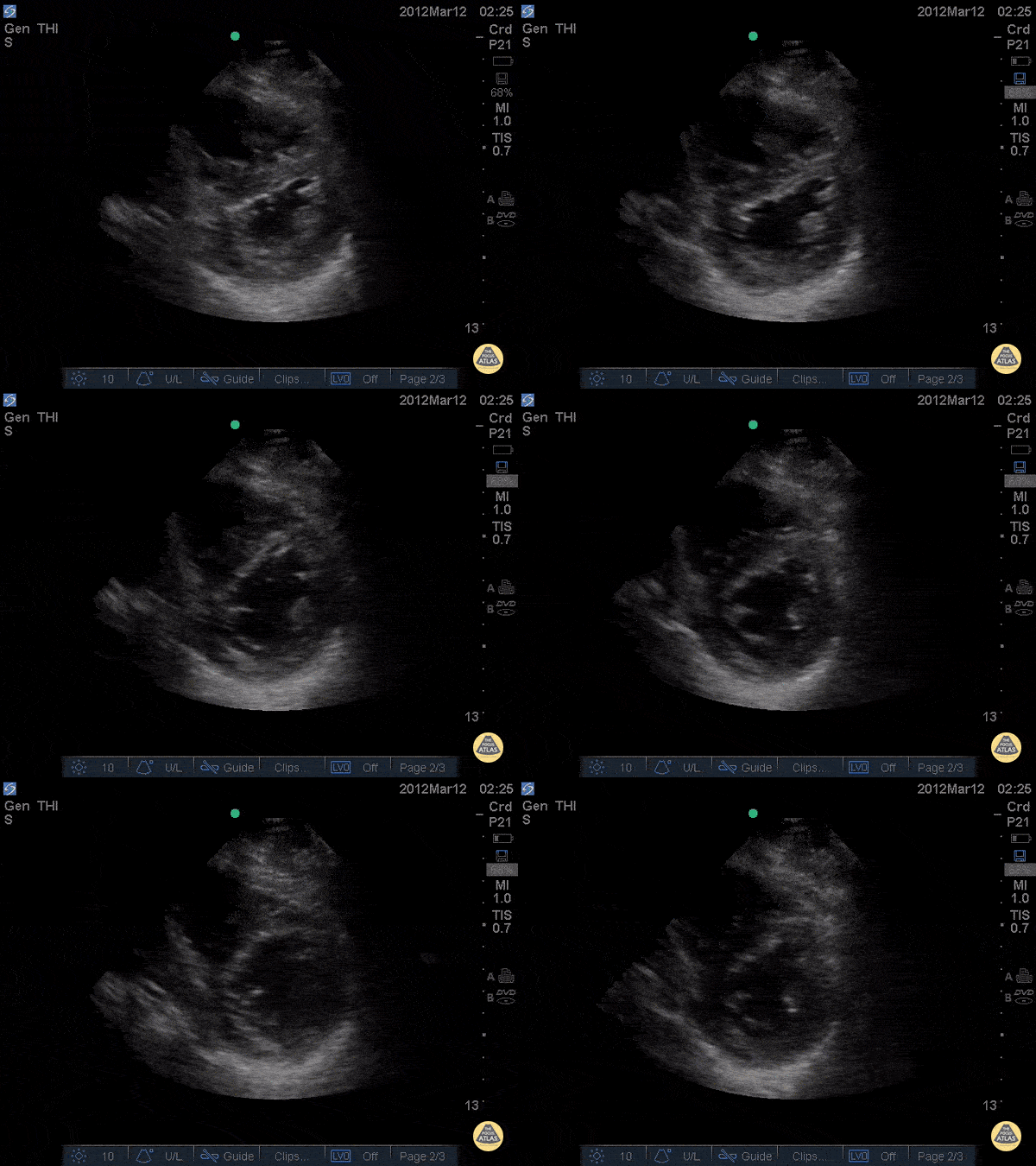
34 y/o female no PMH with chest pain and rapid onset dyspnea on exertion noticed over 1 day. Patient became increasingly tachycardic and ultimately hypotensive so RUSH exam was performed showing D - Sign. CTA was subsequently performed and found to have saddle emboli. Echo was done pre and post TPA showing partial resolution of the D-sign.
Dr. Joshua Schechter - Kings County Emergency Medicine
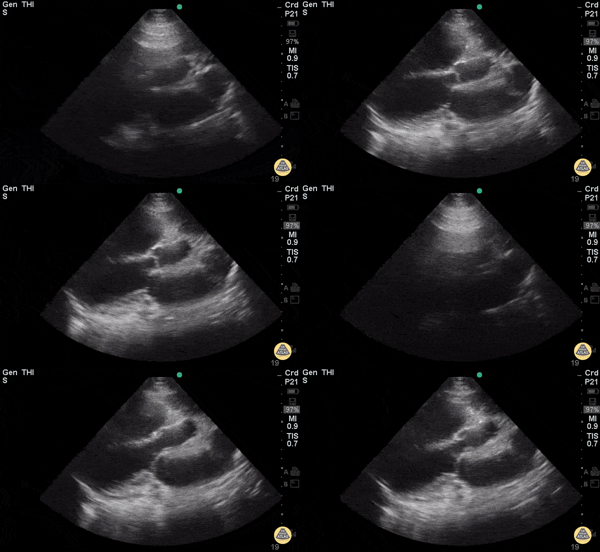
Right-Sided Heart Failure (PLAX)
Young woman with a history of Idiopathic Pulmonary Arterial Hypertension with resultant R heart failure who came in short of breath.
Greg Powell, MD
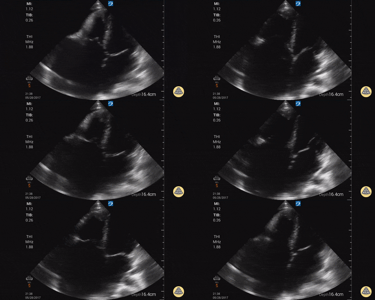
Right-Sided Heart Failure
Young male from Latin America with findings of right HF. Focused cardiac ultrasound shows massive RA, moderate pericardial effusion, relatively normal RV and LV. Significant TR ruled out. Findings consistent with Endomyocardial Fibrosis, found in tropical countries. Need cardiac MRI to confirm. Similar to eosinophilic myocardial fibrosis found in temperate climates.
Dr. Gordon Johnson MD
Internist
Portland Oregon & Uganda
SonoPortlandia.com
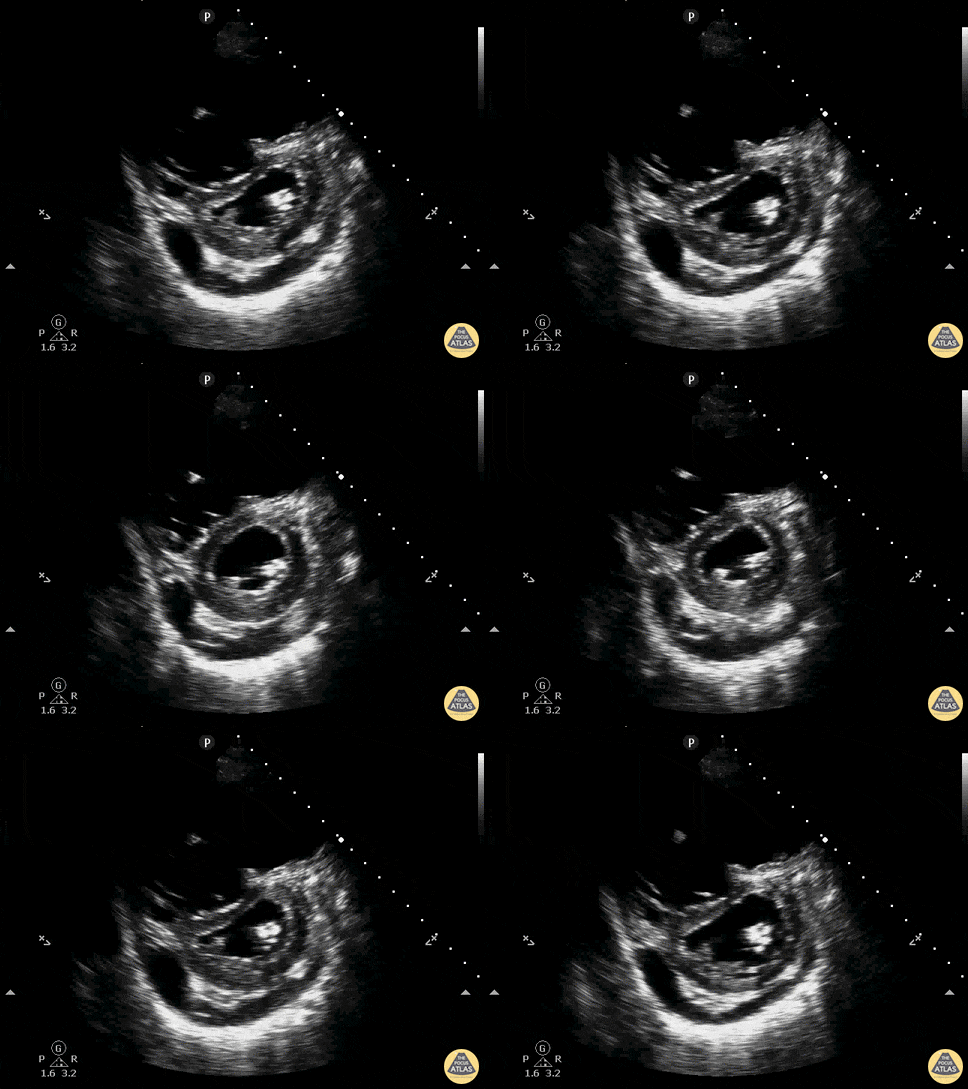
D-Sign with RV Dilation, Effusion, PSSA
A patient presenting with acute onset undifferentiated shortness of breath. POCUS was used to narrow the differential. Parasternal short axis demonstrated flattening of the interventricular septum which pushes the left ventricle into the shape of the letter D. Known as the sonographic D-sign, it is correlated with significant right ventricular overload. This sign is not highly sensitive for PE, but can be 80-90% specific when found and associated with other signs of right ventricular strain. Also note on this study: moderate pericardial effusion, right ventricular dilation.
Drs. Ronald Rivera, Elizabeth Hanson, Melanie Malloy, Kelly Maurelus, Kings County/SUNY Downstate Emergency Medicine
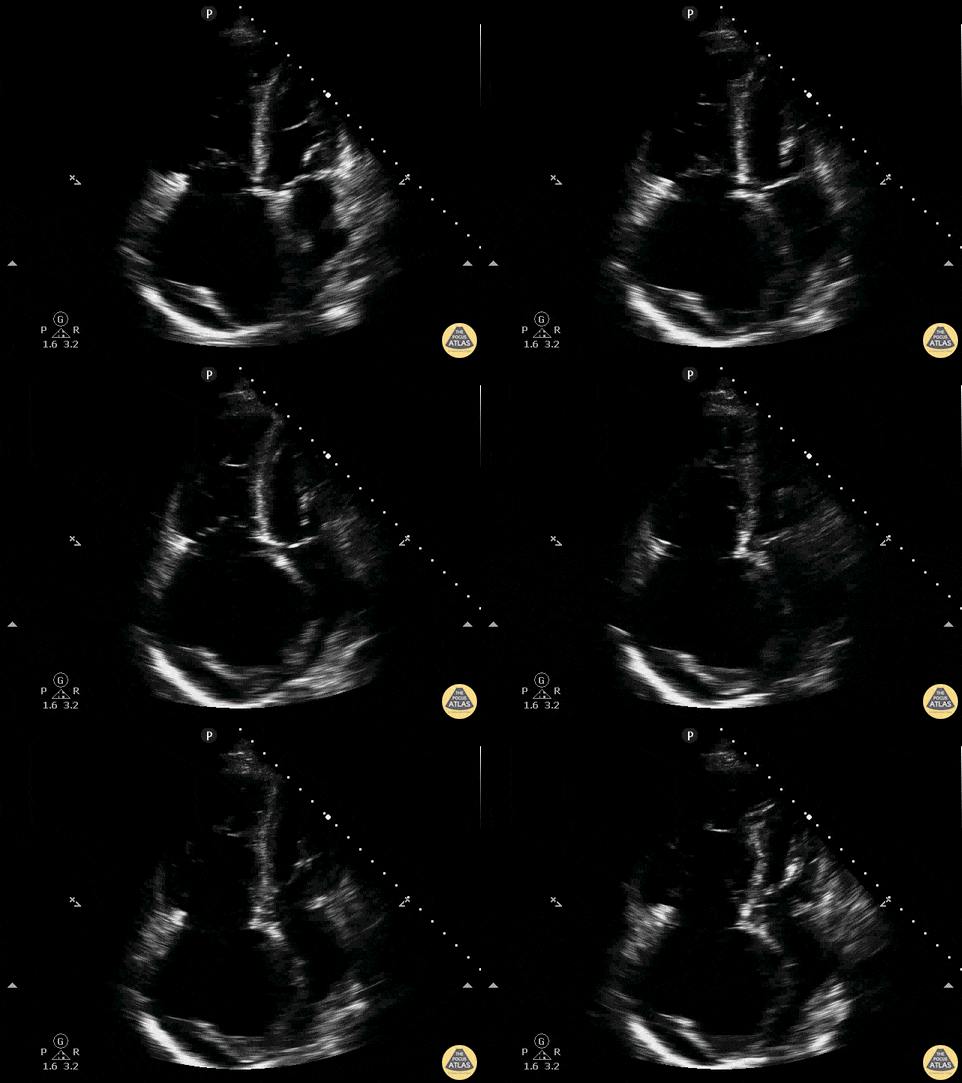
RV Dilation, Effusion, A4C
A patient presenting with acute onset undifferentiated shortness of breath. POCUS was used to narrow the differential. Parasternal short axis demonstrated flattening of the interventricular septum which pushes the left ventricle into the shape of the letter D. Known as the sonographic D-sign, it is correlated with significant right ventricular overload. This sign is not highly sensitive for PE, but can be 80-90% specific when found and associated with other signs of right ventricular strain. Also note on this study: moderate pericardial effusion, right ventricular dilation.
Drs. Ronald Rivera, Elizabeth Hanson, Melanie Malloy - Emergency Medicine Residents
Dr. Kelly Maurelus, Ultrasound Education Director
Kings County/SUNY Downstate Emergency Medicine
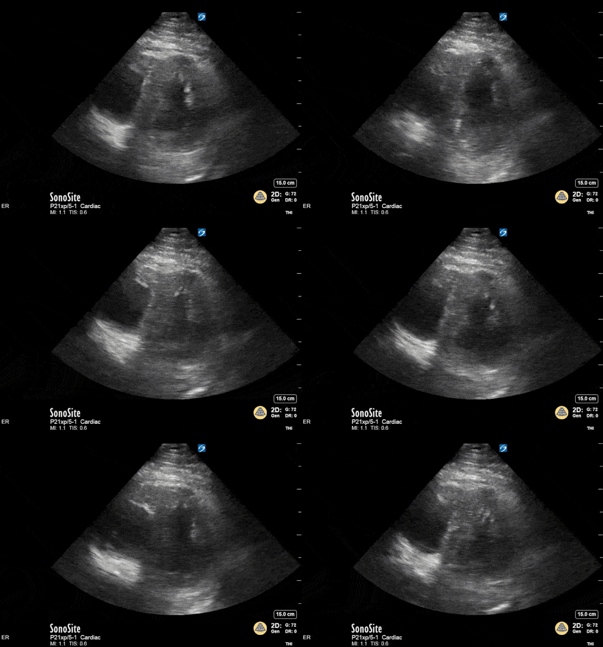
The D Sign
This is a parasternal short axis view in a patient with extensive pulmonary emboli on CT angiogram of the chest. The troponin was mildly elevated and patient hemodynamically stable. A bedside echo revealed evidence of RV strain (note the “D” shaped left ventricle).
Therese Mead, DO
Emergency Physician
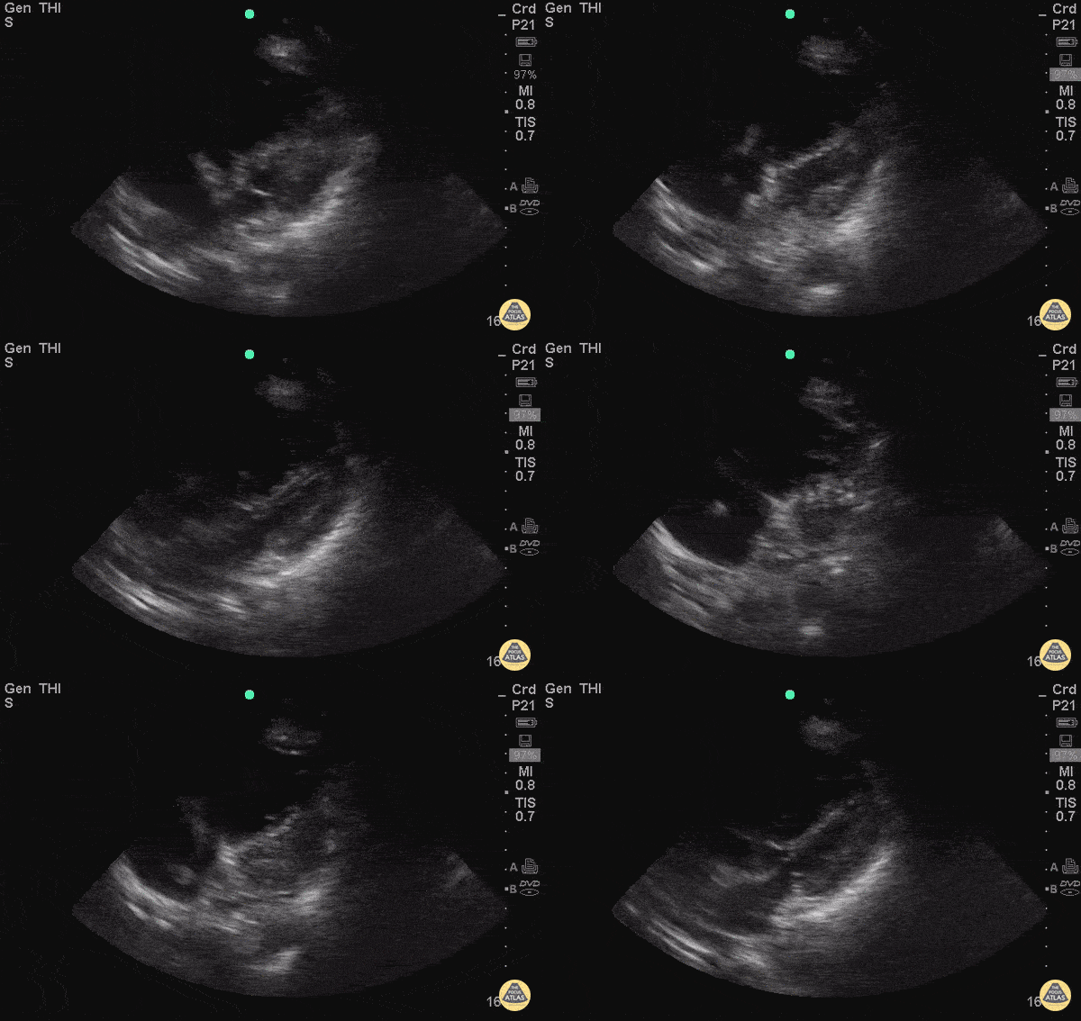
RA Thrombus with R Heart Strain
Middle age F presented initially to ED after a mechanical fall complaining of hip pain. Patient became tachypnic and altered during ER stay. POCUS done, and RA thrombus was noted with D sign and enlarged R ventricle.
R heart thrombus (thrombus in transit) is highly mobile differentiating it from a mural thrombus, which forms in situ. A meta-analysis of 1113 patients from the International Cooperative Pulmonary Embolism Registry showed mortality was 2x as high for pts with right heart thrombus and PE compared to those without right heart thrombus. The difference in mortality was more pronounced in the heparin alone treatment group (vs. lytics or embolectomy).
Another study by Rose et al (2002) showed patients with PE and a right heart thrombus had a mortality of 27%. They found these patients did better when treated more aggressively (i.e. thrombolysis or embolectomy).
Rose PS, Punjabi NM, Pearse DB. Treatment of right heart thromboemboli. Chest. 2002;121(3):806-14. (PMID: 11888964)
Torbicki A, Galié N, Covezzoli A, et al. Right heart thrombi in pulmonary embolism: results from the International Cooperative Pulmonary Embolism Registry. J Am Coll Cardiol. 2003;41(12):2245-51. (PMID: 12821255)
Submitted by Bobak Zonnoor, MD

Acute Cor Pulmonale (Parasternal Long-Axis View)
Justin Bowra MBBS, FACEM, CCPU Emergency Physician, RNSH et al. (Dr. S Fares)
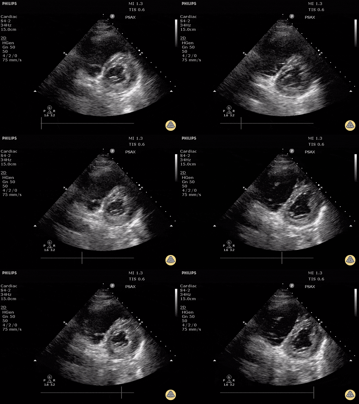
Cor Pulmonale (Short)
Justin Bowra MBBS, FACEM, CCPU Emergency Physician, RNSH et al. (Dr. S Fares)

Cor Pulmonale (Long)
Justin Bowra MBBS, FACEM, CCPU Emergency Physician, RNSH et al.

Right Ventricular Failure with Tricuspid Regurgitation
Significant right ventricular dysfunction with moderate tricuspid regurgitation
Sukh Singh, MD
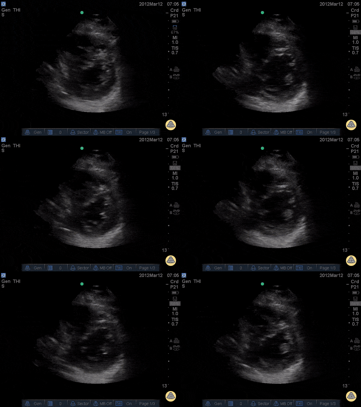
D-Sign in acute PE - Post TPA
34 y/o female no pmh with chest pain and rapid onset dyspnea on exertion noticed over 1 day. Patient became increasingly tachycardic and ultimately hypotensive so RUSH exam was performed showing D - Sign. CTA was subsequently performed and found to have saddle emboli. Echo was done pre and post TPA showing partial resolution of the D-sign.
Dr. Joshua Schechter - Kings County Emergency Medicine
McConnell's Sign
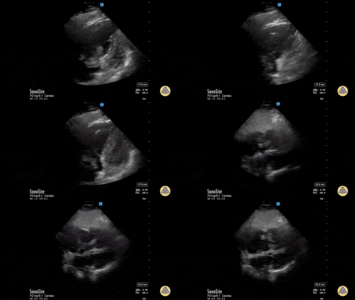
This is a middle-aged male who developed dyspnea suddenly while ambulating. Several weeks prior, he had a laparoscopic procedure with a prolonged post-operative course spanning several weeks in the hospital. He denied chest pain or fever. He was tachypneic with otherwise normal vital signs. Lung sounds were clear, although he had obviously increased work of breathing and appeared diaphoretic. Bedside echocardiography revealed a dilated right ventricle with a positive McConnell sign. There was also a large, lobulated, and mobile hyperechoeic mass within the right atrium suspicious for thrombus. CTA of the chest showed bilateral pulmonary emboli. BNP and troponin were moderately elevated, consistent with submassive pulmonary embolism.
Andrew Goodrich, MS, DO, PGY-3
Chief Resident, Central Michigan University Emergency Medicine Residency
Therese Mead, DO, RDMS, FACEP
Associate Program Director and Ultrasound Director, Central Michigan University Emergency Medicine Residency
Right Atrial Thrombus
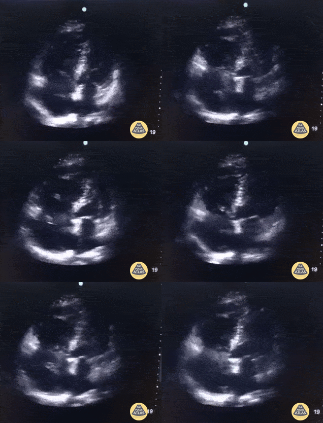
A 70 year old female patient with complaints of acute dyspnea. Oxygen saturation 86% with room-air improved with O2-suppletion. Poor hemodynamics with systolic presure around 100 mmHg. POCUS shows obvious right heart strain with apical sparing (McConnel's sign) and trombus drifting around in right atrium and even hitting the right ventricle. Correct therapy (trombolytics) was started almost straight away after presentation at the ED because POCUS was available.
Dr. Van Roosmalen
Acute PE with McConnell's Sign
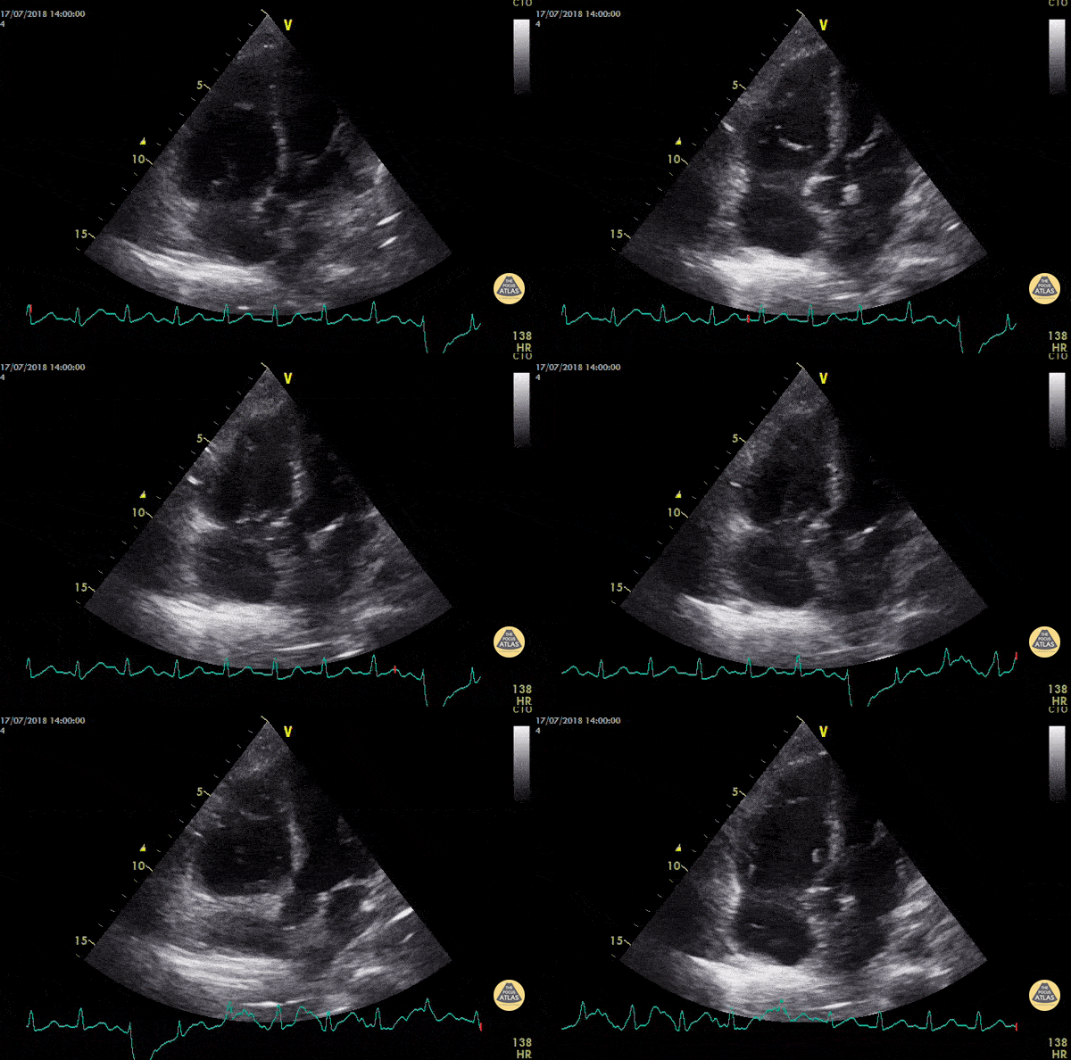
46 year old man, intraoperative cardiac arrest after massive pulmonary embolism. Apical four chamber view obtained after resuscitation showed McConnell's sign. We found other signs of acute cor pulmonale : the 60/60 sign and RV dilatation.
Dr. Devigne

Clot in Transit & Femoral DVT
A 73 year old male presented with several day history of moderate dyspnea as well as right LE discomfort. POCUS identified both a right intracavitary thrombus as well as right femoral phlebitis. He was treated with heparin for both.
Pierre Bernatas, @pb2316
Emergency Physician. Limousin, France.

Right Atrial Thrombus
Elderly female with hypotension and shortness of breath and back pain presenting in extremis. Treated with TPA for a RA thrombus and saddle pulmonary embolism.
Dr. William Scheels

RV Dysfunction and Pulmonary Hypertension from COPD
50s M with PMH COPD presented with dyspnea and increasing oxygen requirement. POCUS is shown here, demonstrating significant RV dilation, reduced RV function, and bowing of the septum concerning for pulmonary hypertension. Additional workup ultimately ruled out ACS and DVT/PE, and the patient was admitted for COPD exacerbation, and formal TTE confirmed a new diagnosis of pulmonary hypertension.
Dr. Cailin Frank, Ultrasound Fellow, Denver Health Ultrasound Fellowship
Dr. Anna Engeln, Denver Health Medical Center

Pulmonary Hypertension from Triscupid Endocarditis
30s F with PMH IVDU and prior endocarditis s/p tricuspid valve repair presented with fever, hypotension, and dyspnea. On RUSH evaluation, this cardiac image was acquired, showing significant RV dilation with bowing of the septum into the LV. The prosthetic tricuspid valve is shown here, but without color doppler, evaluation of the valve patency is limited. Formal TTE showed a patent tricuspid valve however with moderate to severe tricuspid regurgitation, likely explaining the severe RV overload.
Dr. Michael Duerson, PGY4
Denver Health Residency in Emergency Medicine


