Thoracic Aortic Dissection & Aneurysm

Aortic Dissection Flap in Arch of Aorta
A 65-year-old male presents with shortness of breath (no chest pain) and was found to have a dilated aortic root on CT pulmonary angiogram. POCUS (supra sternal view) showed a dissection flap in the arch of aorta; a finding subsequently confirmed on CT aortagram. Patient was sent for emergency surgical intervention.
Dr.Rajasutharsan Kathirgamanathan, Emergency Physician
The Northern Hospital, Melbourne, Australia
@raj_kathir007

Suprasternal View of Type A Dissection
Suprasternal notch view shows a mobile intimal dissection flap in the aortic arch.
Michael Cover, MD
@michaelc0ver

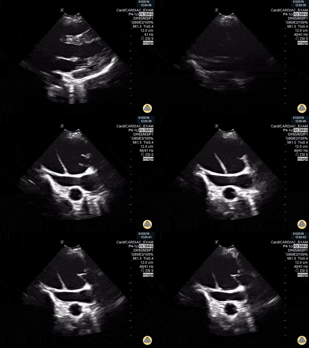
Type A Aortic Dissection - Double Valve Sign
Type A aortic dissection flap appears as a "second valve" on this parasternal long axis cardiac view.
Michael Cover, MD
@michaelc0ver

Descending Thoracic Aortic Dissection Seen on PSLA
This is a parasternal long axis view of an elderly male with PMH of hypertension and DM presenting with a dissection of the descending aorta (aka type B aortic dissection).
Image courtesy of Robert Jones, DO, FACEP @RJonesSonoEM
Director, Emergency Ultrasound; MetroHealth Medical Center; Professor, Case Western Reserve Medical School, Cleveland, OH
Find his original post here

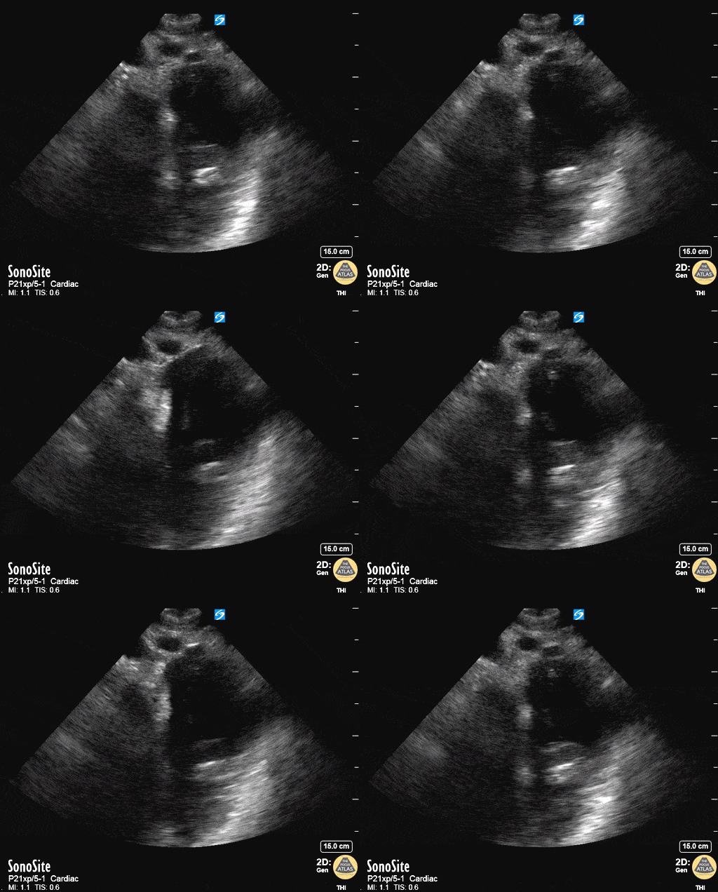
Descending Thoracic Aorta Flap Seen on Apical 4 Chamber
33 yo male presented with chest/epigastric pain. POCUS is notable for acute aortic dissection as seen in the descending thoracic aorta flap as well as presence of a pericardial effusion.
Maxime Gautier

Aortic Dissection Flap Visualized in Proximal Aorta with Root Dilation
This is a parasternal long axis view demonstrating significant enlargement of the aortic root with an identified dissection flap located in the proximal ascending aorta.
Frances Russell, MD, RDMS
Assistant Professor of Emergency Medicine Division Chief, Ultrasound Fellowship Director, Ultrasound

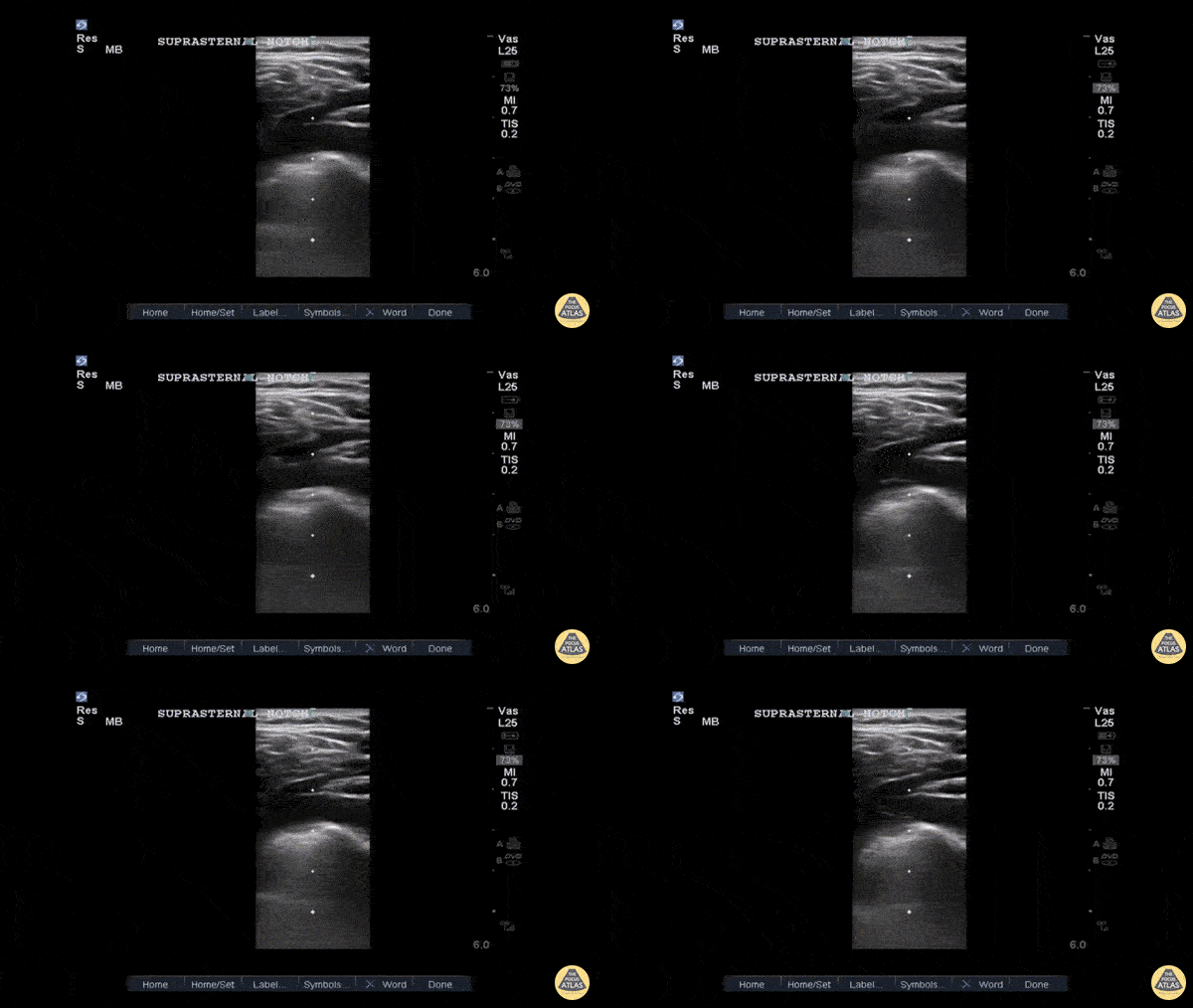
Type B Thoracic Aortic Dissection Flap on Suprasternal View
This is a suprasternal notch view demonstrating an aortic flap in a patient with a Stanford Type B thoracic aortic dissection.
This 40ish year old was a truck driver with untreated hypertension with sudden onset interscapular pain that migrated to his lumbar area. He stopped, lost strength in his right leg and was transported to our ED. The POCUS allowed the CV surgeon to prepare while the confirmatory CTA and standard treatment were performed. Suprasternal notch imaging with the linear or fine parts probe in a patient with suspicious signs/symptoms allows for a more rapid diagnosis of thoracic aortic dissection.
John E. Hipskind, MD, FACEP
Clerkship Director
ED, Kaweah Delta Hospital

Aortic Dissection On Suprasternal View
50 year old mandarin speaking man complains of vague central chest pain. You pursue a routine cardiac workup which is fairly normal. Upon discharging him, the nurse tells you his systolic in now in the 80's.
After clenching the likely diagnosis of an Type A Aortic Dissection based on your echo, you confirm by placing the transducer in the patients suprasternal notch, transversely but with the probe marker rotated slightly toward the patient's hip and fan inferiorly. You see a grossly widened aorta and a dissection flap.
Dr. Matthew Riscinti - Kings County Emergency Medicine, Dr. Benjamin Clearly - NYU Langone Emergency Medicine


