
Valvulopathy

Mitral Valve Vegetation
32-year-old woman presented to ED with clinical signs of subacute stroke - confirmed via Brain CT . In the investigation of etiology of cerebral injury, POCUS identified this large hyperechoic vegetation on the mitral valve (seen here both in PLAX and PSAX views). As a result of these images, a diagnosis of infectious endocarditis causing cerebral septic emboli injury was considered.
Renato Tambelli; @JediPocus
Emergency Physician (HCFAMEMA /Sao Paulo, Brazil)

Mitral Regurgitation in Parasternal Long Axis
Parasternal long axis view in a patient with severe mitral regurgitation.
Rohan Rastogi, MD
@RohanRastogiMD

Tricuspid Regurgitation
A patient with IVC and hepatic congestion on CT also had a holosystolic murmur at the left lower sternal border, worsened with inhalation. POCUS revealed severe tricuspid regurgitation on parasternal short axis view with color doppler.
Image courtesy of Robert Jones DO, FACEP @RJonesSonoEM
Director, Emergency Ultrasound; MetroHealth Medical Center; Professor, Case Western Reserve Medical School, Cleveland, OH
View his original post here

Tricuspid Valve Endocarditis
Tricuspid valve endocarditis seen in a patient with IVDU. This subcostal view allows great visualization of the tricuspid valve when using the liver as an acoustic window.
Image courtesy of Robert Jones DO, FACEP @RJonesSonoEM
Director, Emergency Ultrasound; MetroHealth Medical Center; Professor, Case Western Reserve Medical School, Cleveland, OH
View his original post here

Mitral Valve Endocarditis
A patient presented to the ED with fever, sepsis, Janeway lesions, Osler nodes, and splinter hemorrhages. PLAX view revealed a vegetation on the mitral valve indicative of MV endocarditis.
Image courtesy of Robert Jones DO, FACEP @RJonesSonoEM
Director, Emergency Ultrasound; MetroHealth Medical Center; Professor, Case Western Reserve Medical School, Cleveland, OH
View his original post here

Functional MR in Heart Failure
A 58-year-old male with ischemic cardiomyopathy and recently implanted AICD presented with subacute dyspnea without signs of volume overload on physical exam. Seen here is POCUS (apical four chamber view) notable for mitral valve regurgitation (MR) as demonstrated by the presence of a regurgitant jet. After excluding acute myocardial ischemia, patient was diagnosed with secondary (functional) MR due to heart failure.
Separately, notice the hyperechoic lesion traversing the right atrium. In this patient it represents a segment of his AICD. The differential diagnosis, however, includes atrial thrombus and/or vegetation.
Shahad Al Chalaby, MD. PGY3 Internal Medicine Highland Hospital. Alemeda Health System Internal Medicine Residency Program. CA, USA
@shahad_Chalaby
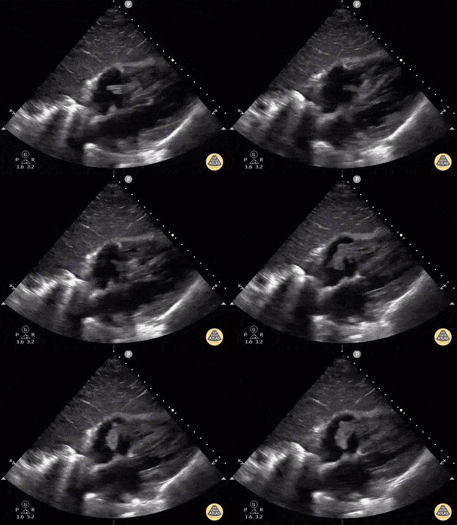
Tricuspid Endocarditis
Subcostal view of a patient with a history of IV drug use. Ultimately this patient was diagnosed with a polymicrobial endocarditis of the tricuspid valve with MRSA and Candida Albicans as culprits. Note the hyperechoic lesion swinging between the right atrium and right ventricle.
Image courtesy of Robert Jones DO, FACEP @RJonesSonoEM
Director, Emergency Ultrasound; MetroHealth Medical Center; Professor, Case Western Reserve Medical School, Cleveland, OH
View his original post here
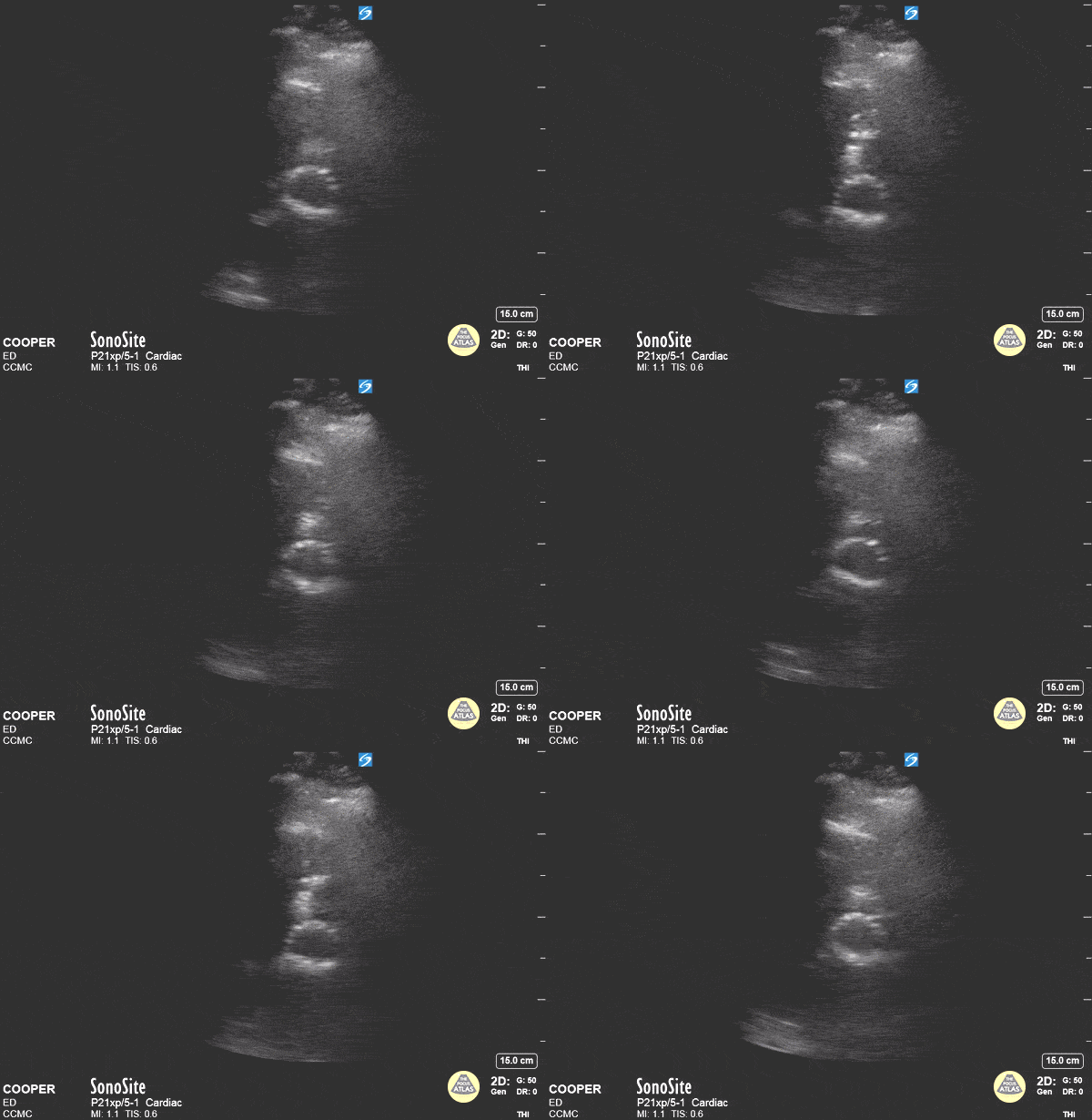
TAVR in Short Axis
A 71-year-old male was brought to the ED via EMS for exertional chest pain. The patient was a poor historian and vaguely described a prior cardiac stenting procedure. Bedside echocardiography was performed and demonstrated a hyperechoic ring-shaped structure at the cardiac base when viewed in the parasternal short axis. This structure was determined to be a Transcatheter Aortic Valve Replacement (TAVR). This finding improved the care teams understanding of the patients medical history and altered their evaluation to include pathology of the proximal aorta.
Arthur Gross, DO; Max Cooper, MD - Crozer-Chester EM

MV Inflow Variation
This patient presented with acute blood loss anemia and high output heart failure. Note the significant mitral valve inflow respiratory variation secondary to hypovolemia (not attributed to the trace pericardial effusion). This nicely illustrates potential mitral/tricuspid valve inflow variation in hypovolemic patients.
Luka Petrovic, Chief Medical Resident Rutgers New Jersey Medical School
@lukapetrovic89
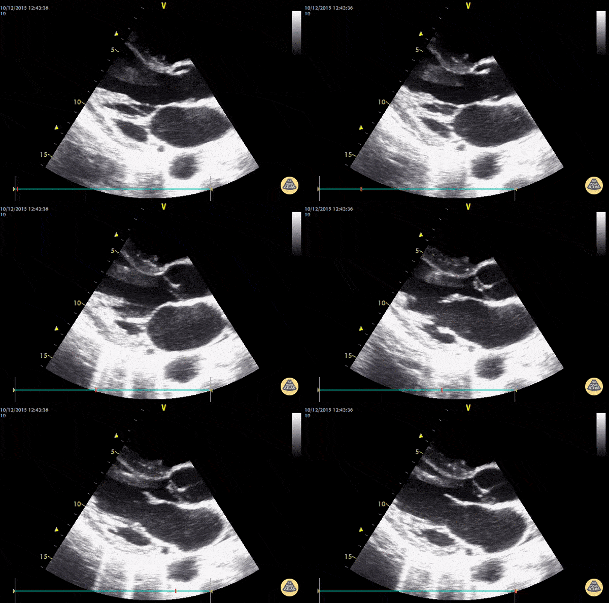
Endocarditis
Vegetations on the AV valve (endocarditis). 40y old female coming to the ED with acute ischemia of the left lower limb and fever. IVDA.
Dr. Dominik Doeller
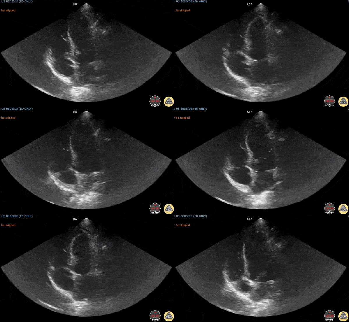
Endocarditis in an Intravenous Drug User
A careful look will show something extra on the mitral valve.
Dr. Sarah Banks, Dr. Molly Thiessen - Denver Health Emergency Medicine

Severe Mitral Regurgitation
60s M with PMH ischemic cardiomyopathy and CHF presented with multiple episodes of syncope. The initial workup was unrevealing so POCUS was performed. These color doppler images demonstrate severe mitral regurgitation as seen as a multicolored retrograde jet from the mitral valve during systole. This patient was admitted for further workup of his syncope and telemetry monitoring given the structural heart disease and concern for underlying dysrhythmia.
Dr. Nhu-Nguyen Le, Fellow
Denver Health Ultrasound Fellowship
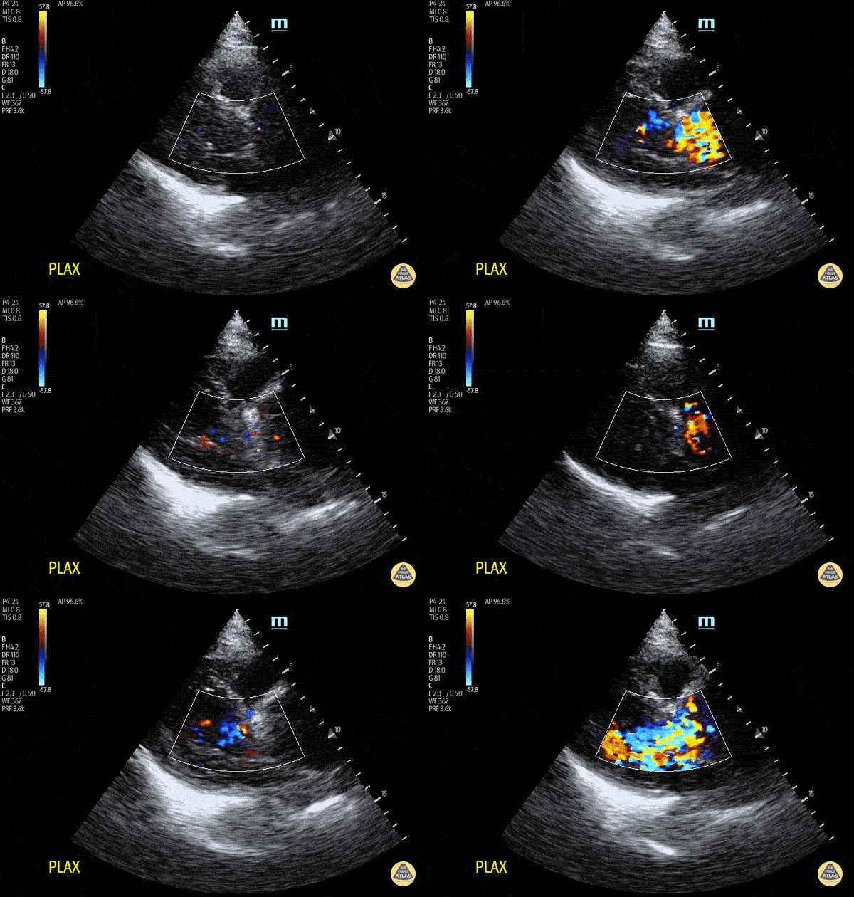
Aortic Root Dilation with Aortic Regurgitation
This images demonstrates a dilated aortic root in a patient with chest pain radiating to the back. Note that the ratio of the RV outflow tract, aortic root, and left atrium are not 1:1:1. Whilst you may not always see an intimal flap, a dilated aortic root and new aortic regurgitation may indicate acute aortic dissection. In this case, this was subsequently confirmed on CT aortogram.
Dr. Peter Cheng
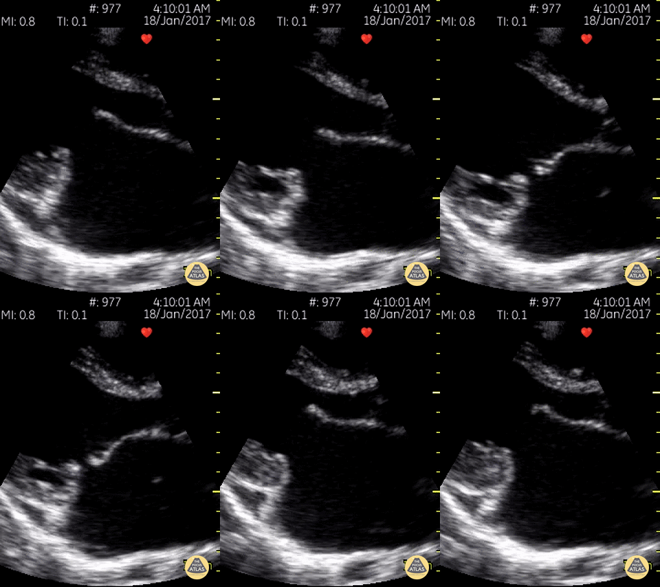
Hockey Stick Sign
Mitral stenosis with mitral regurgitation with the classic MV "hockey stick" sign of rheumatic mitral valve disease.
Dr. Gordon Johnson MD
Internist
Portland Oregon & Uganda
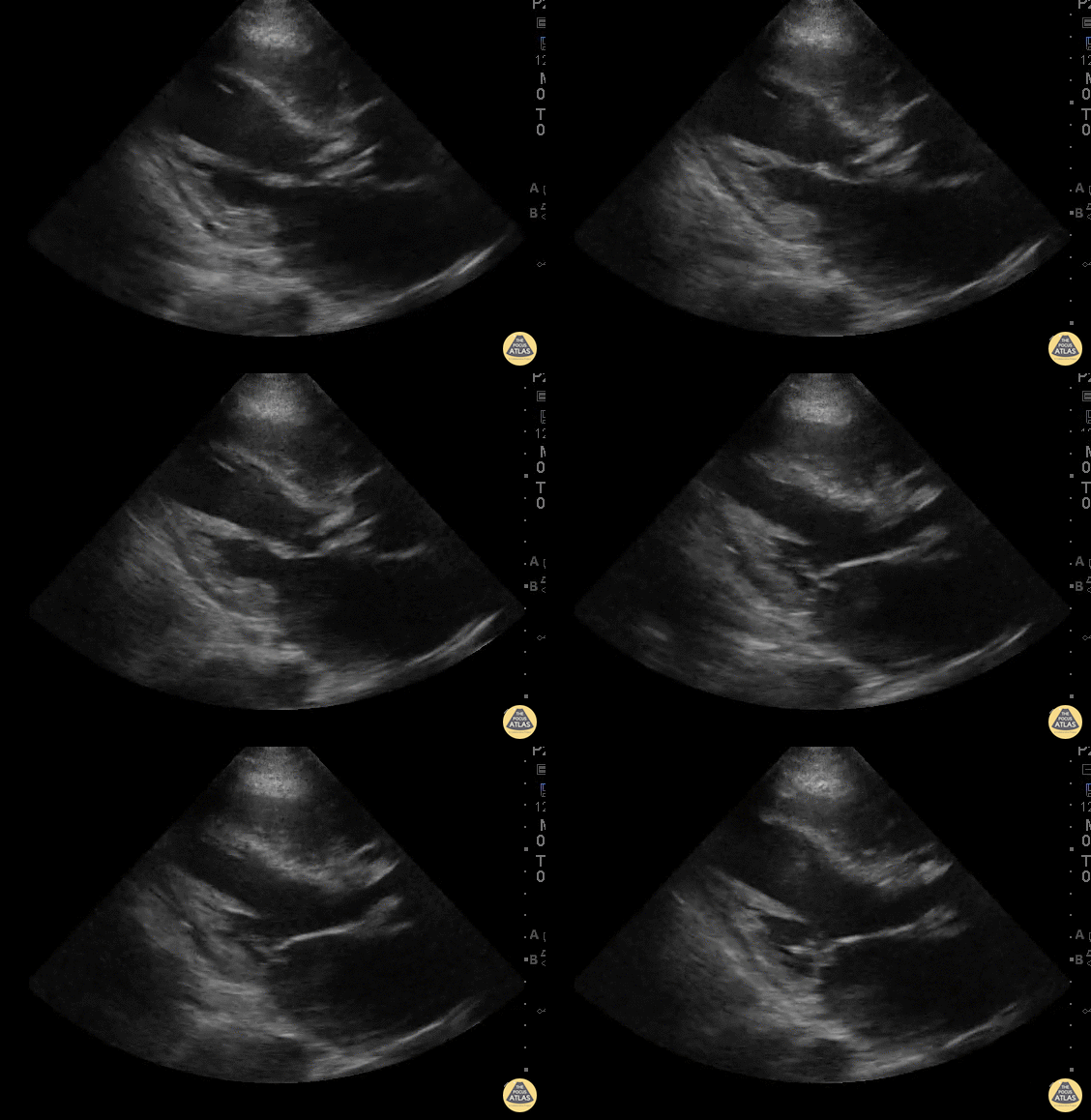
Aortic & Mitral Valve Vegetations
WCUME 2017 Submission for "Best POCUS"
40 year old male who presented with 1 week low grade fever and mild pedal edema and orthopnea.
POCUS demonstrates a small vegetation on the anterior leaflet of the mitral valve as well as large vegetations on the aortic valve with obvious poor coaptation of valve leaflets.
Dr. Peh Wee Ming - Singapore General Hospital
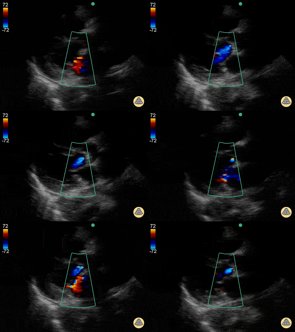
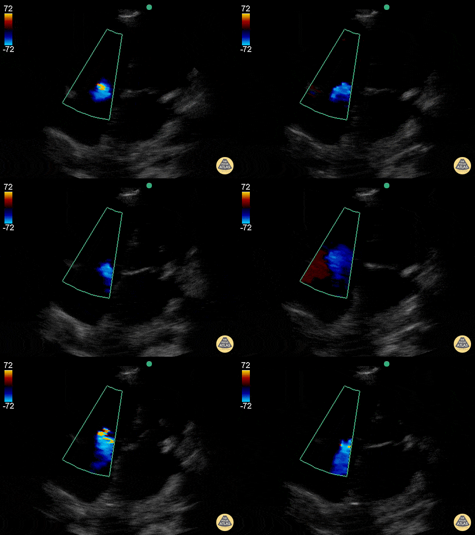
Aortic and Mitral Regurgitation
This was a patient who presented at the age of 98 who had become progressively more short of breath over the last several months and now had trouble getting around. Very sharp and witty woman, who wished to have no aggressive measures. She was tucked into the cardiology service for gentle diuresis and optimization of her heart disease. This parasternal long axis demonstrating alternating mild-moderate aortic regurgitation with moderate mitral regurgitation.
Jason Tanguay, DO
Mild Tricuspid Regurgitation
A narrow, central tricuspid regurgitation jet is seen on this apical 4-chamber view consistent with mild tricuspid regurgitation.
Jason Tanguay, DO
Moderate Mitral Regurgitation
A mitral regurgitant jet is seen in this apical 4-chamber view that appears to be ~35-40% the area of the left atrium. This is most consistent with moderate MR though a more quantitative method such as PISA can be used for formal evaluation.
Jason Tanguay, DO
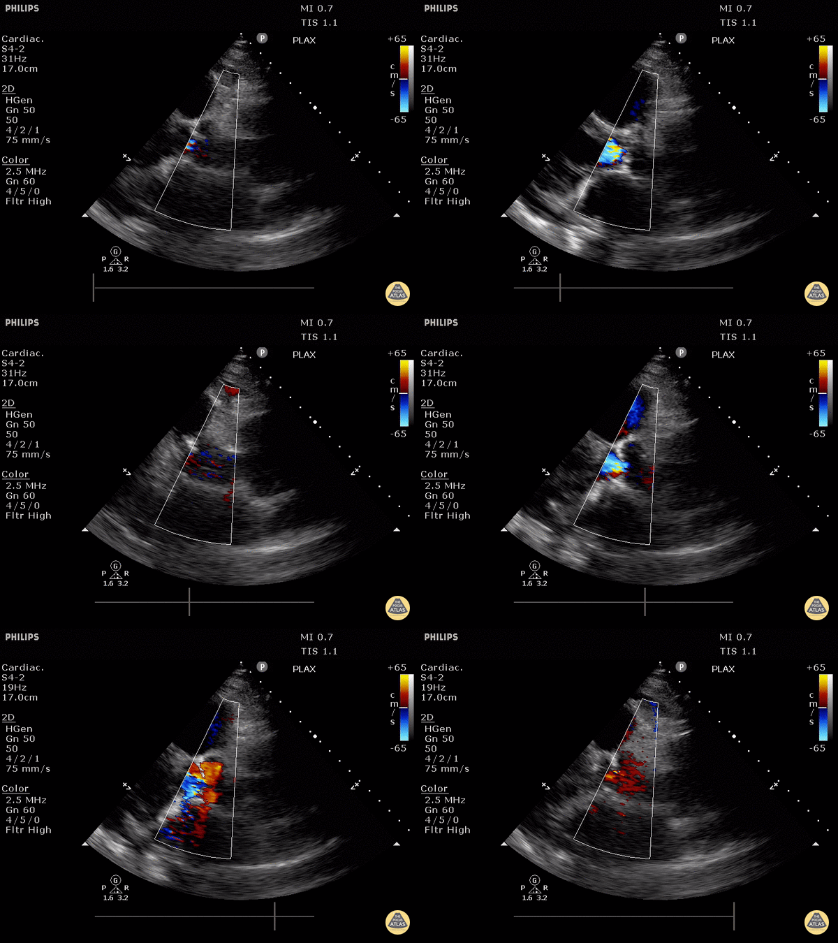
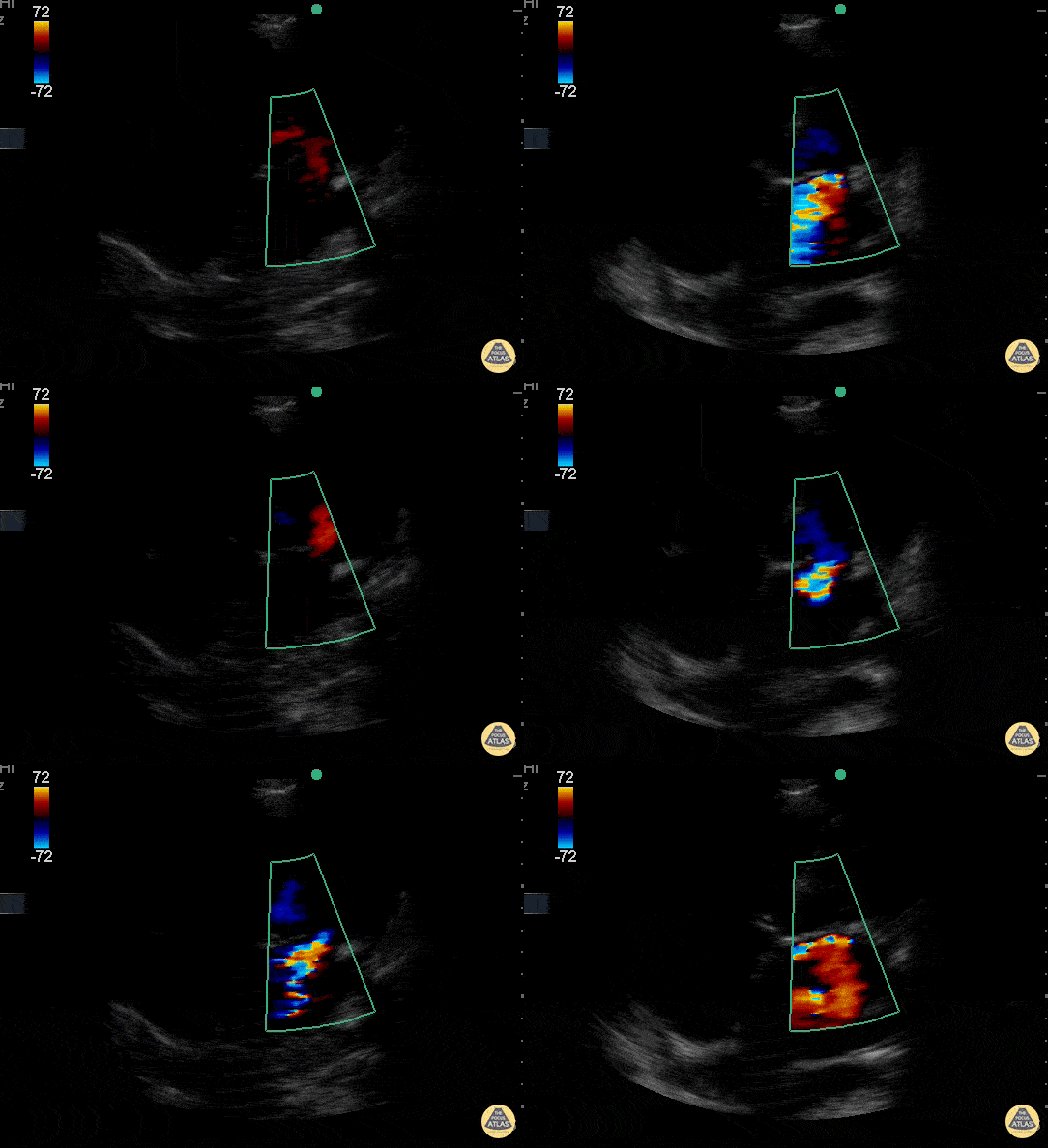
Moderate Aortic Regurgitation
Justin Bowra MBBS, FACEM, CCPU Emergency Physician, RNSH et al. (Dr. Giles and Dr. Jacob)
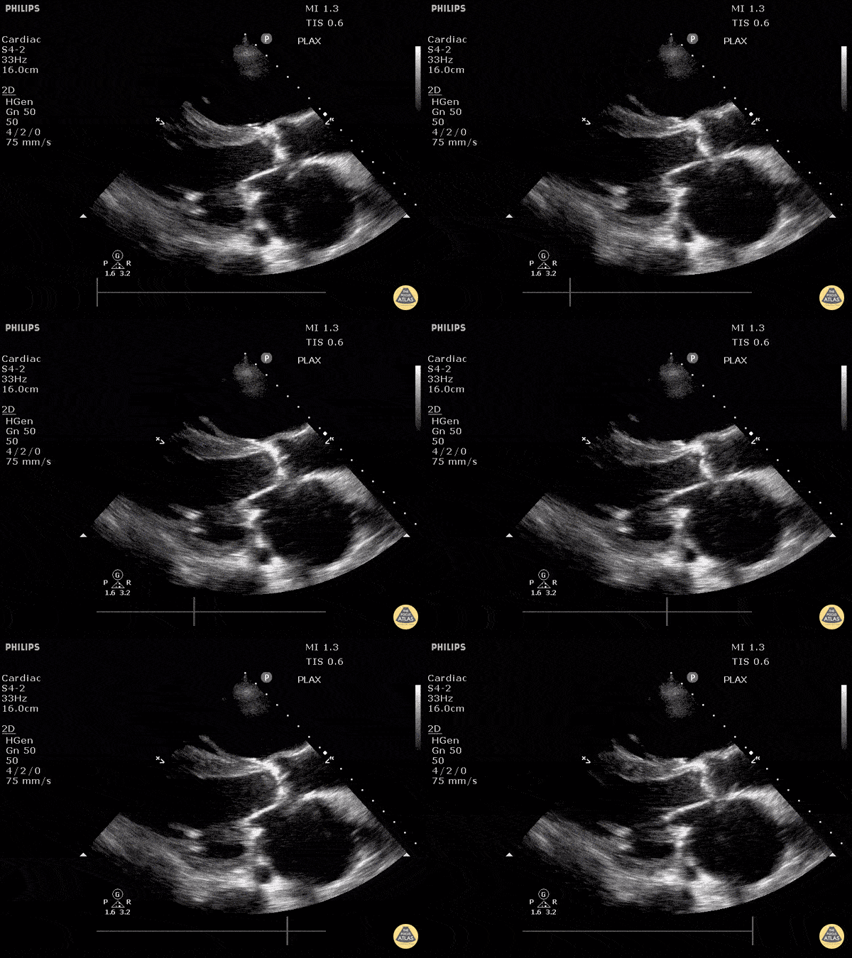
Severe Aortic Stenosis
Justin Bowra MBBS, FACEM, CCPU Emergency Physician, RNSH et al.
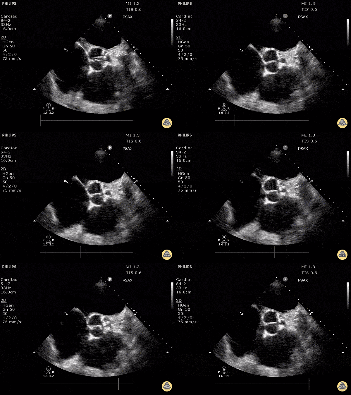
Severe Aortic Stenosis
Justin Bowra MBBS, FACEM, CCPU Emergency Physician, RNSH et al.
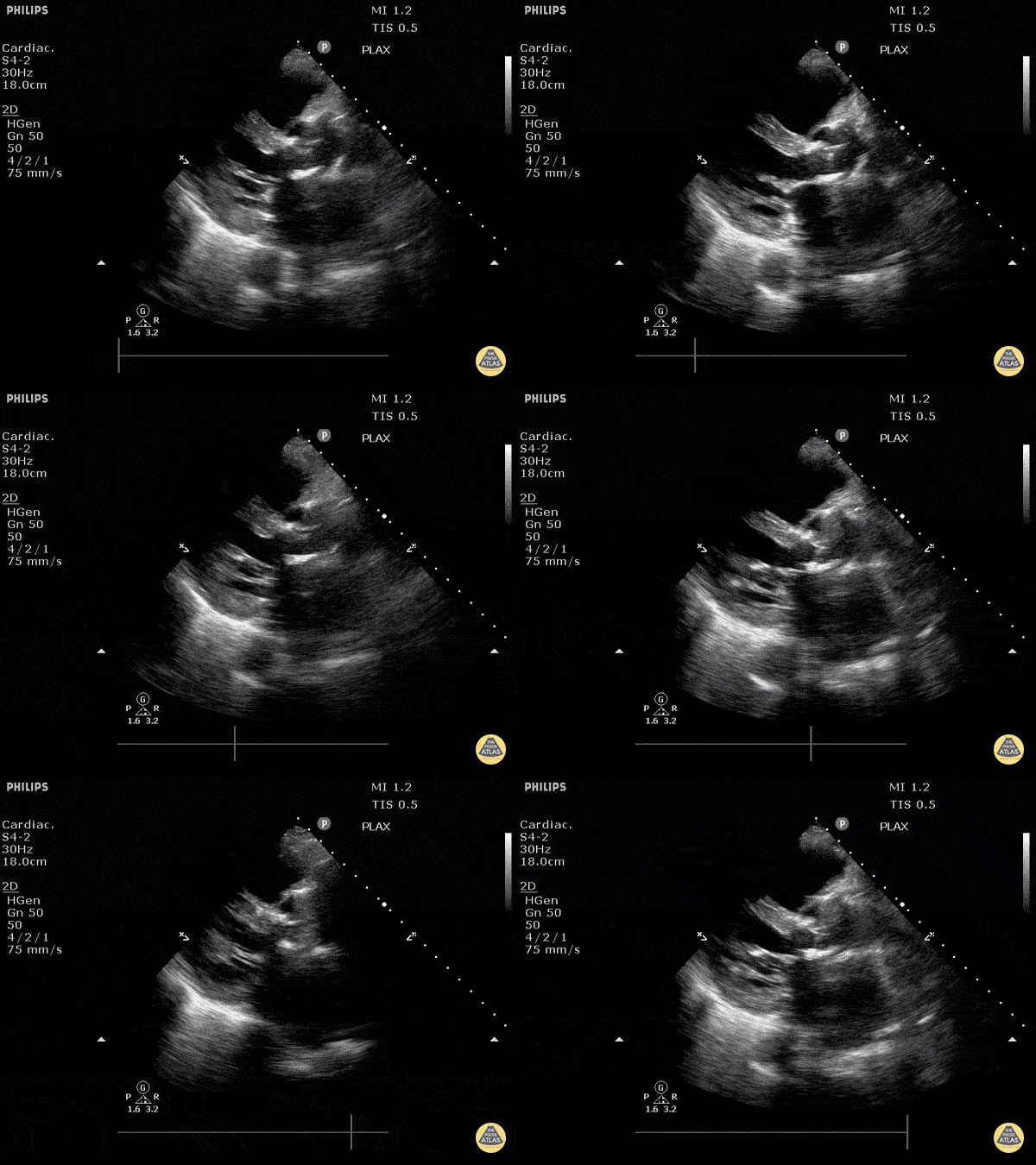
Aortic Valve Prosthesis (Long)
Justin Bowra MBBS, FACEM, CCPU Emergency Physician, RNSH et al. (with Dr. Orr)
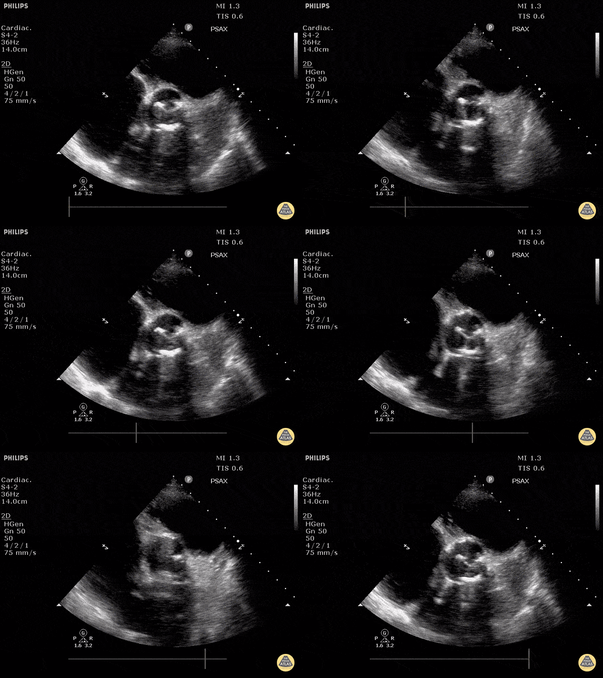
Aortic Valve Prosthesis (Short)
Justin Bowra MBBS, FACEM, CCPU Emergency Physician, RNSH et al. (with Dr. Orr)
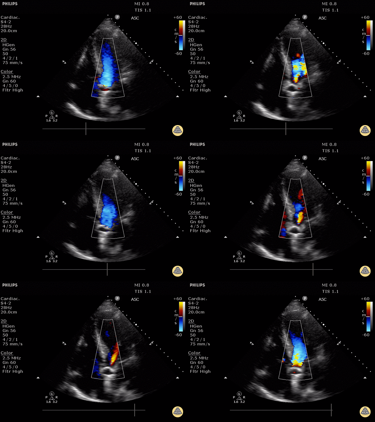
Mild Aortic Valve Regurgitation
Justin Bowra MBBS, FACEM, CCPU Emergency Physician, RNSH et al. (Dr. Mo Haywood)
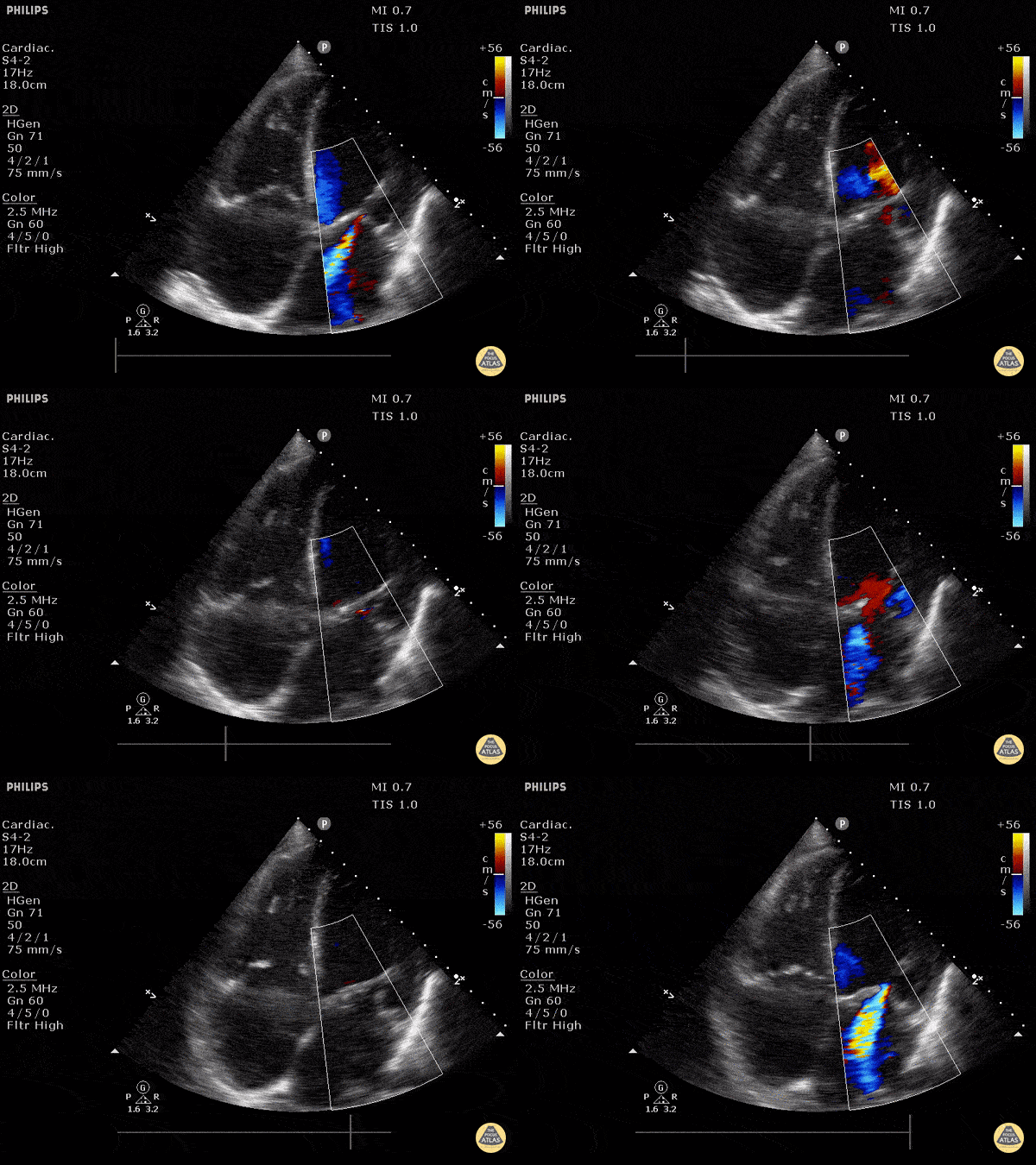
Heart Failure with Moderate Mitral Regurgitation
Justin Bowra MBBS, FACEM, CCPU Emergency Physician, RNSH et al. (Dr. K Kaynama)
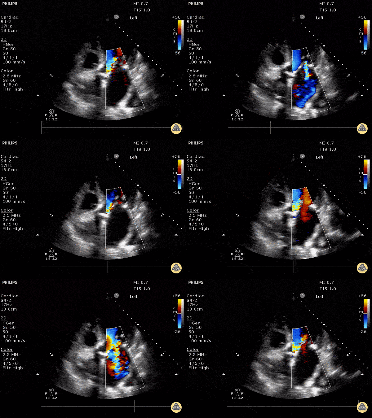
Mixed Aortic and Mitral Regurgitation
Mixed aortic and mitral regurgitation with color doppler.
Justin Bowra MBBS, FACEM, CCPU Emergency Physician, RNSH et al.
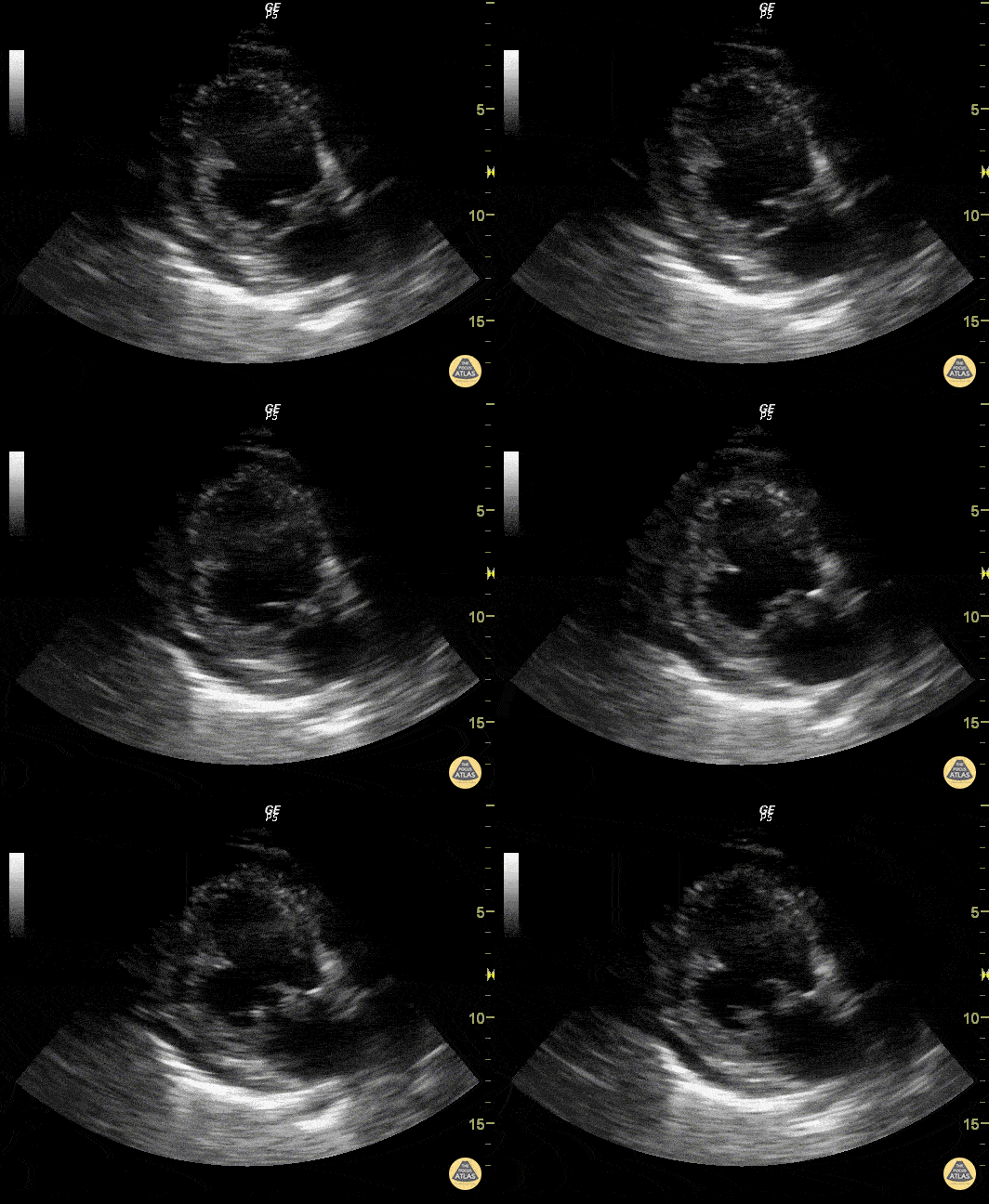
Mitral Valve Prolapse (Apical Long-Axis View)
Justin Bowra MBBS, FACEM, CCPU Emergency Physician, RNSH et al. (Dr. Pankaj Arora)
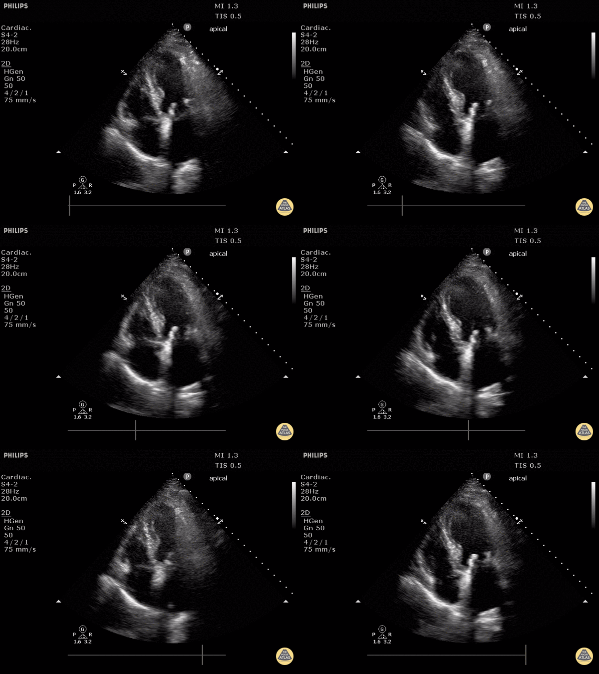
Prosthetic Mitral Valve (Apical 4 Chamber View)
Justin Bowra MBBS, FACEM, CCPU Emergency Physician, RNSH et al.
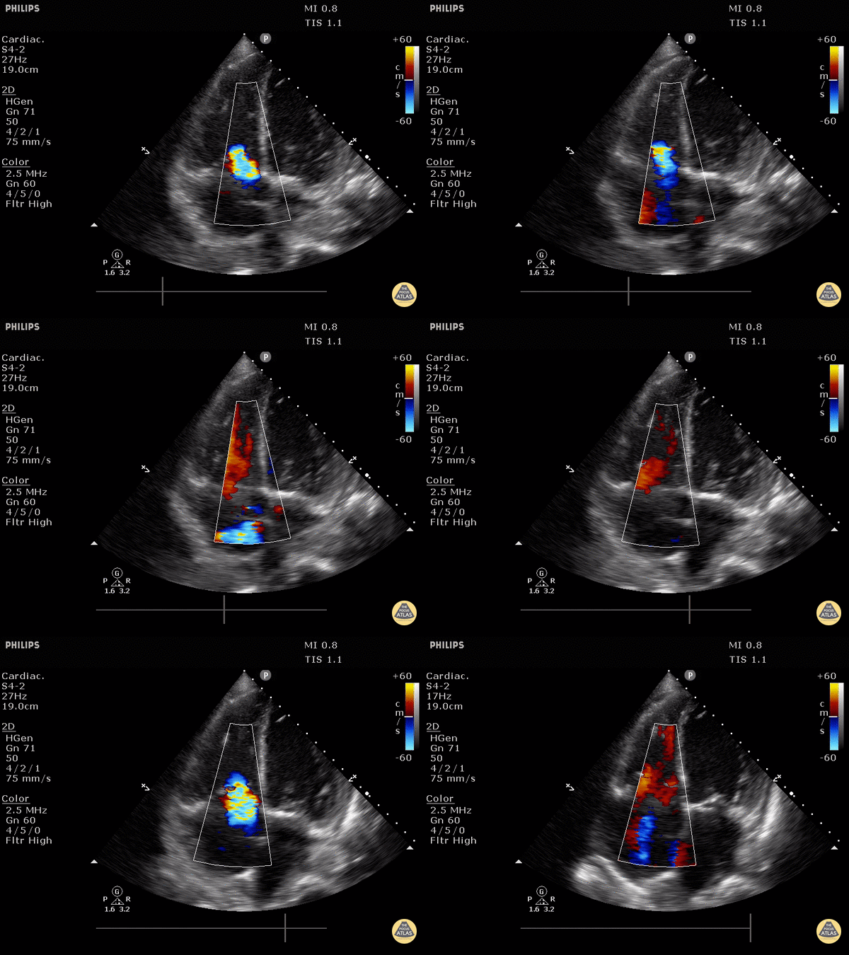
Tricuspid Valve Regurgitation
Moderate to severe tricuspid regurgitation demonstrated on apical 4 chamber view.
Justin Bowra MBBS, FACEM, CCPU Emergency Physician, RNSH et al. (Dr. Kaynama)
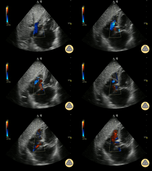
Tricuspid Valve Regurgitation
Subcostal View demonstrating mild tricuspid regurgitation
Justin Bowra MBBS, FACEM, CCPU Emergency Physician, RNSH et al.

Mitral Regurgitation
Moderate mitral regurgitation
Sukh Singh, MD
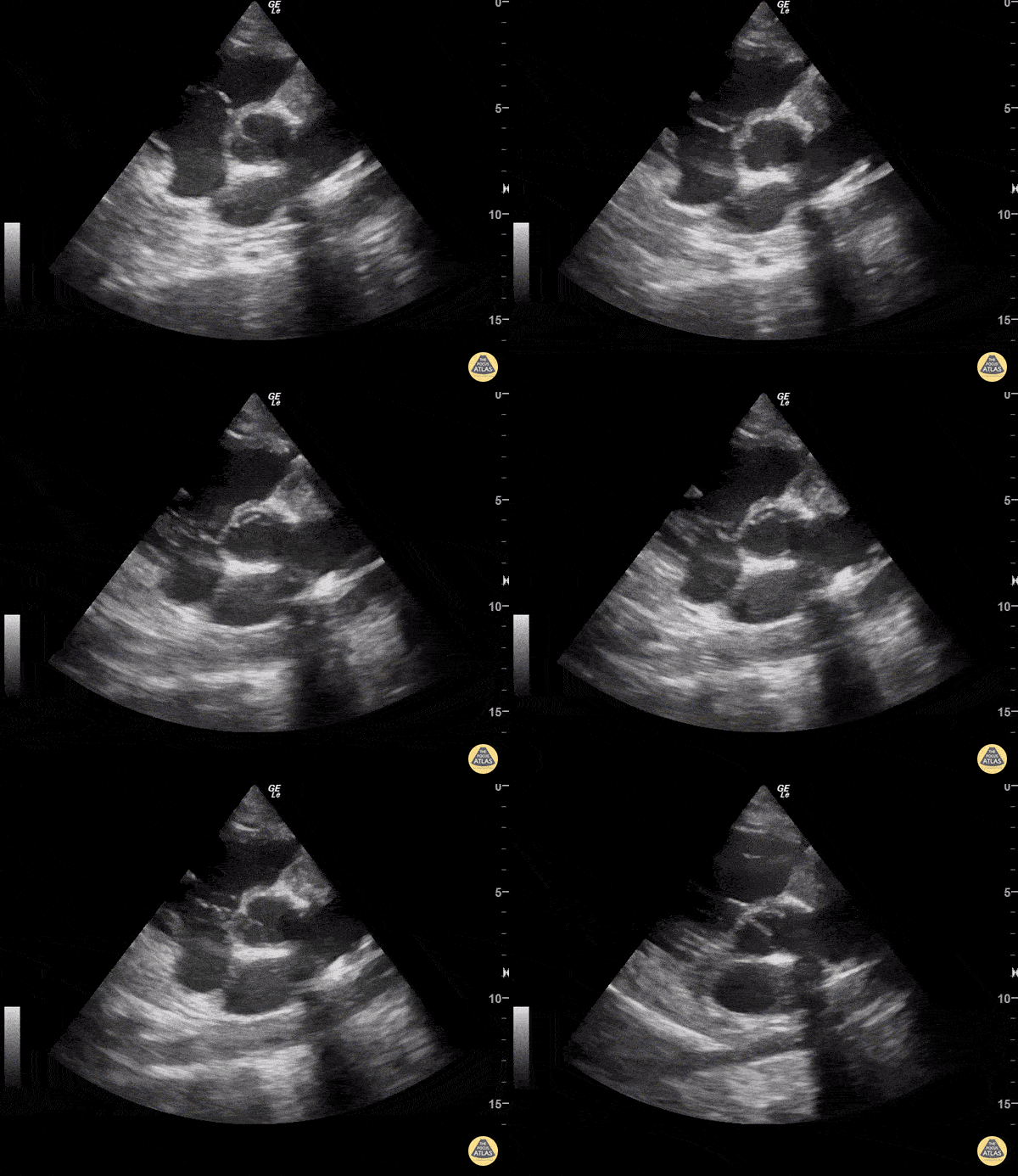
Pulmonic Vegetation
Sukh Singh, MD
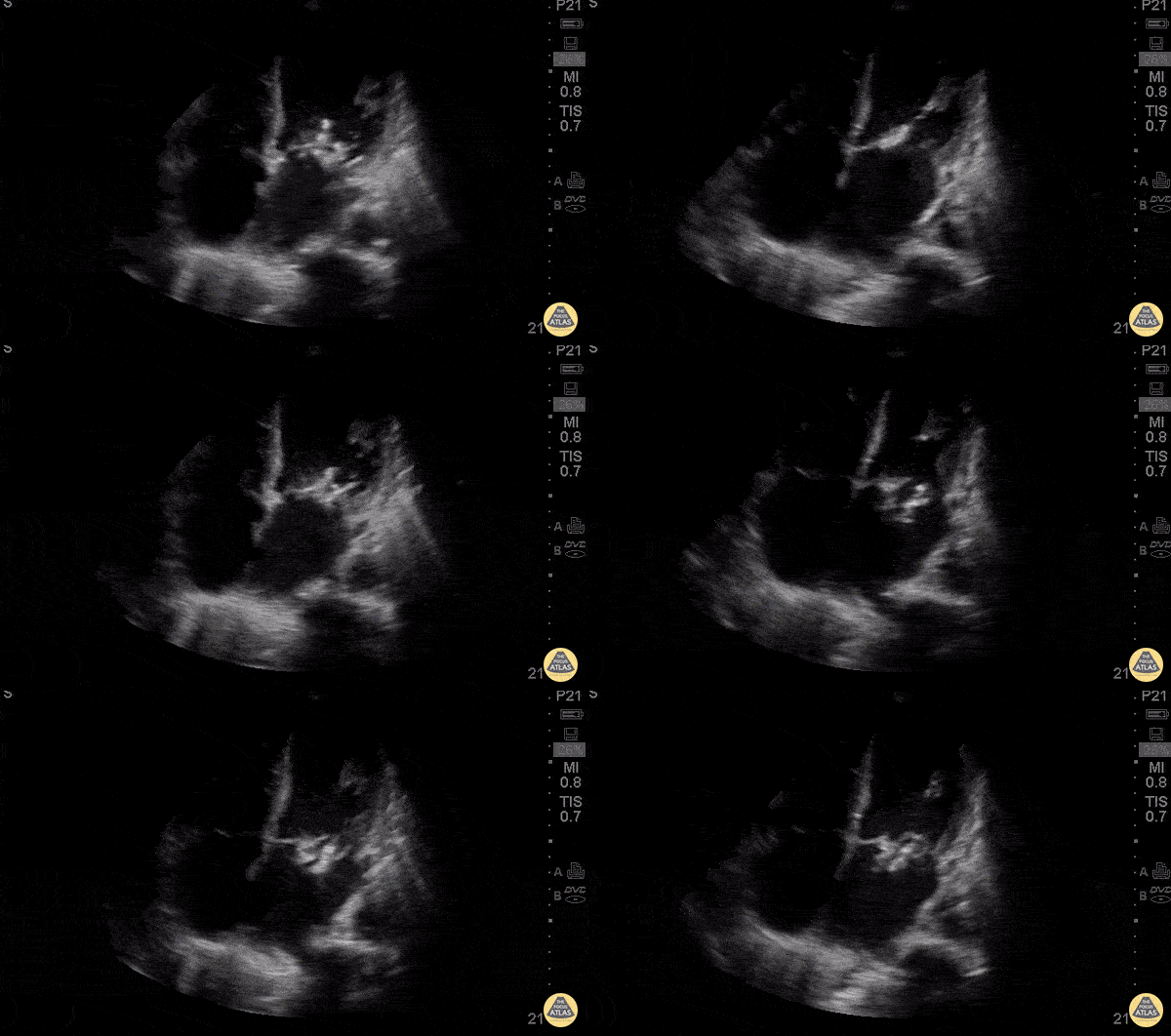
Mitral Vegetations
62 y/o M no PMH visiting from the Caribbean presents with 3 days of acute onset shortness of breath associated with severe exertional dyspnea, orthopnea and cough productive of whitish sputum without fevers or chest pain.
POCUS was performed to evaluate undifferentiated shortness of breath. Subcostal view revealed vegetations on both mitral valve leaflets with severe mitral regurgitation, biatrial enlargement and global hypokinesis. Acute onset of severe heart failure requires emergent surgical intervention including mitral valve repair vs replacement. In our patient, POCUS lead to emergent transfer for CT surgery.
Dr’s Karen Benabou, Eden Kim, and Eric Schnitzer - Kings County Emergency Medicine

Tricuspid Valve Vegetation
50-60s F with PMH IVDU presents with chest pain, dyspnea, and fever. This POCUS image shows the parasternal short axis view at the level of the aortic valve, revealing a large vegetation on the tricuspid valve. Color doppler demonstrates tricuspid regurgitation.
Daniel Fuchs, PGY3
Denver Health Residency in Emergency Medicine

Mitral Clip
A 54-year-old man with history of dilated cardiomyopathy on maximum tolerated doses of guideline directed medical therapy and implantable cardioverter defibrillator presented with progressively worsening dyspnea with minimal activity. Transthoracic ultrasonography showed evidence of severe functional mitral regurgitation for which he underwent mitral clip placement (seen here). This point of care ultrasound was obtained during a clinic follow up when he reported sustained resolution of symptoms; there was also no appreciable mitral regurgitant jet on color doppler.
Shahad Al Chalaby, MD (PGY3) @shahad_Chalaby
Alameda Health System Internal Medicine Residency Program
Oakland, California

Infective Endocarditis
A 35-year-old with history of a "heart problem" presented for concern of COVID-19. He was found to be in wide complex tachycardia with hemodynamic instability (MAP 50) with associated findings of crushing chest pain and diaphoresis. RUSH demonstrated severely reduced EF, a tricuspid valve vegetation, and aortic valve vegetation complicated by aortic root abscess. Seen here is a parasternal short axis view (on the left) demonstrating an almost entirely occluded aortic valve and outflow tract. The apical four chamber view (on the right) demonstrates both a tricuspid valve vegetation as well as this patient’s severely reduced LVEF.
Gregory Wiener, MD. Denver Health Residency in Emergency Medicine
@DenverEMed

Left Ventricular Outflow Tract Obstruction
Septic patient with hypotension shows LVOT obstruction with systolic anterior motion of the mitral valve on echo.
Image courtesy of Robert Jones DO, FACEP @RJonesSonoEM
Director, Emergency Ultrasound; MetroHealth Medical Center; Professor, Case Western Reserve Medical School, Cleveland, OH
View his original post here
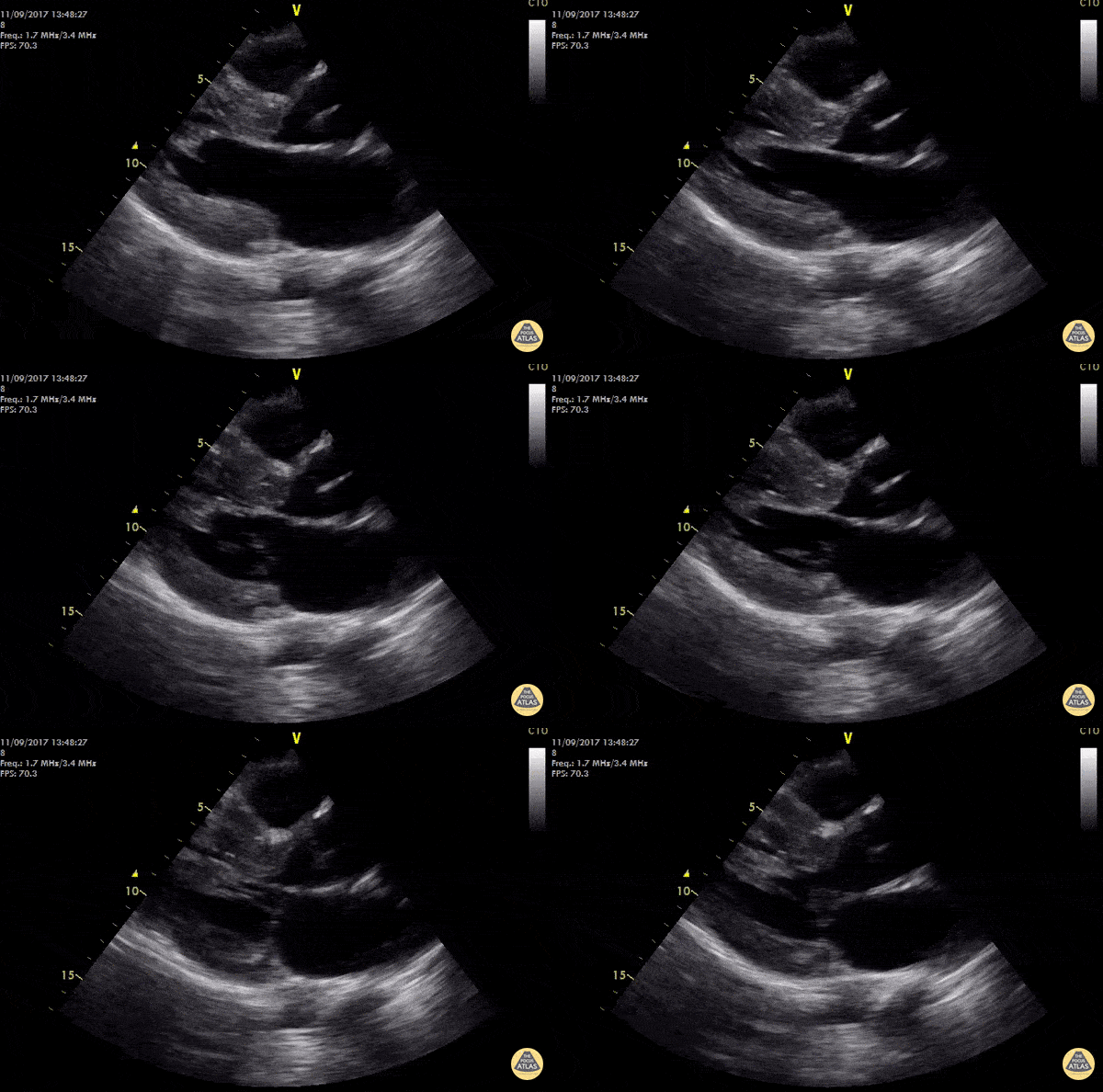
Hypertrophic Obstructive Cardiomyopathy
WCUME 2017 Submission for "Creative Caption" - "Uncle SAM"
19 year old male out of hospital cardiac arrest s/p ROSC. POCUS shows hypertrophic interventricular septum with systolic anterior motion of the mitral valve (SAM) causing LVOT obstruction.
LVOT gradient was measured at 118 mmHg and AICD was fitted during hospital stay. Treatment for HCM usually recommended if: SAM lesion visualized, IVS >18mmm, LVOT gradient > 30mmHg.
Cian McDermott, MD
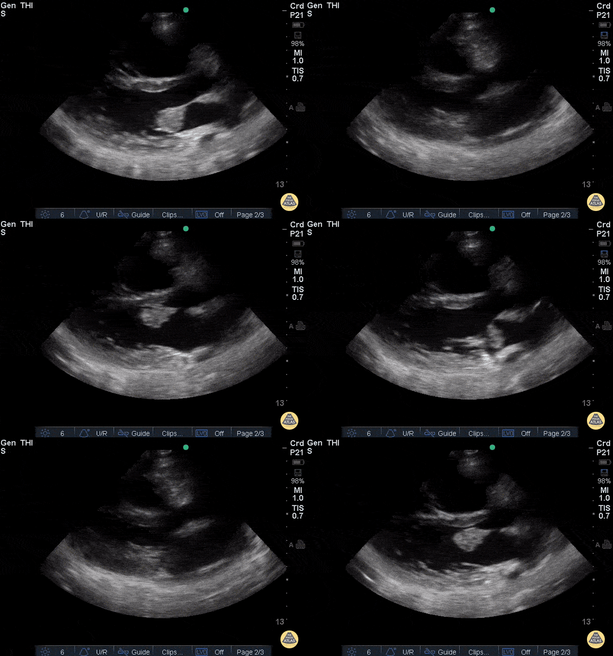
Endocarditis PLAX
Patient with history of IV drug use, admitted for sepsis. Parasternal long axis view shows large mass attached to anterior leaflet of mitral valve. Blood cultures prove bacterial endocarditis.
Ria Dancel, MD. University of North Carolina at Chapel Hill
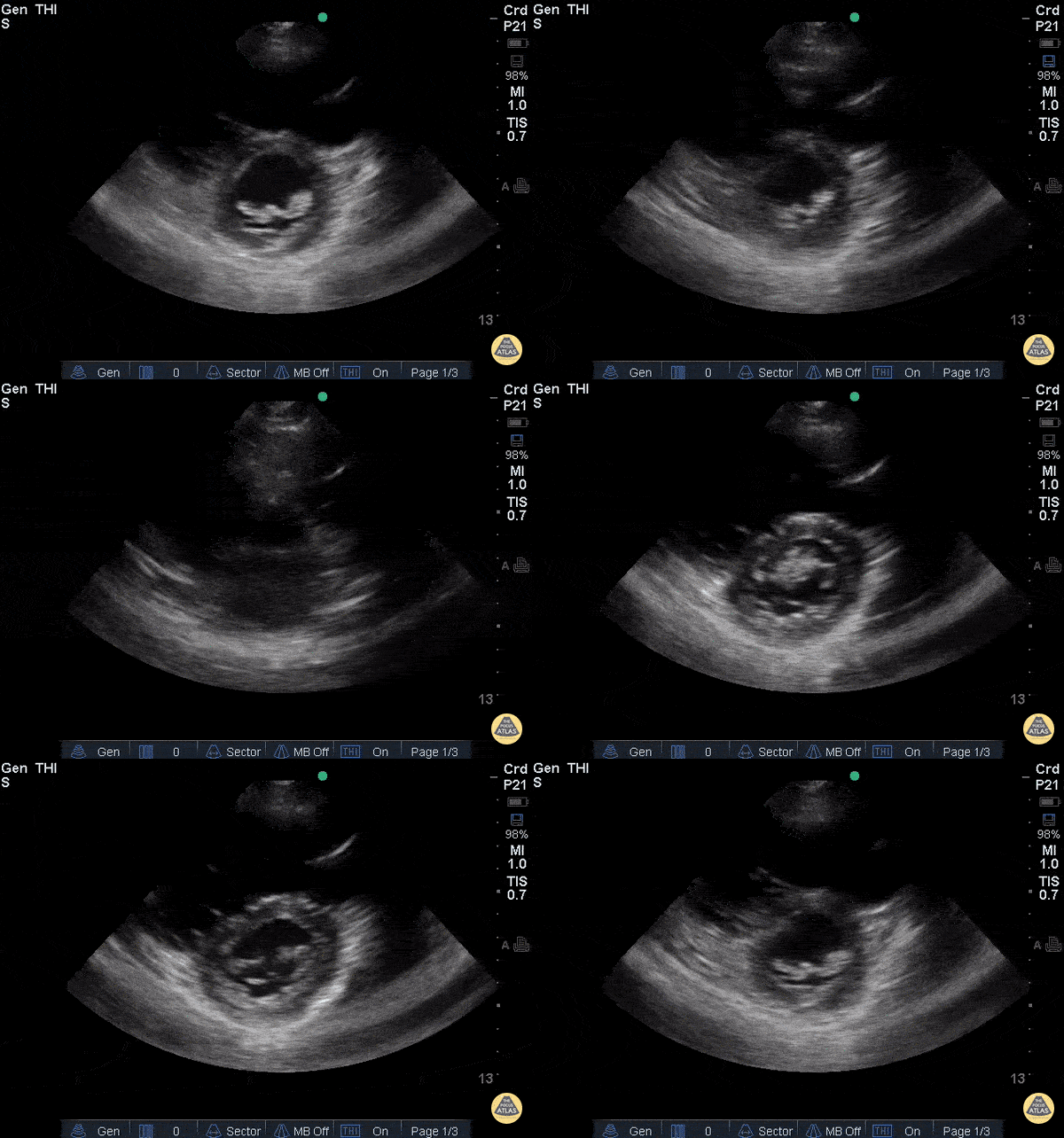
Endocarditis PSAX
Patient with history of IV drug use, admitted for sepsis. Parasternal short axis view shows large mass attached to anterior leaflet of mitral valve. Blood cultures prove bacterial endocarditis.
Ria Dancel, MD. University of North Carolina at Chapel Hill


