
IVC & Abnormal Venous Waveforms

Clot At Junction of Right Atrium and IVC
This patient initially presented post-operatively to the emergency department with complaints of dyspnea. As we fan through this saggital view of the IVC as it enters the right atrium, we can see hyperechoic structures suggestive of clot formation. An alternative view of this clot from a subxiphoid view can be seen here. The patient was subsequently diagnosed with a DVT that extended into their central femoral vein, at the same leg that was recently operated on.
Image courtesy of Robert Jones DO, FACEP @RJonesSonoEM
Director, Emergency Ultrasound; MetroHealth Medical Center; Professor, Case Western Reserve Medical School, Cleveland, OH
View his original post here
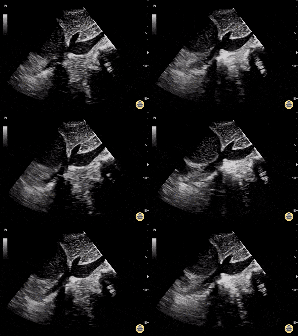
Look at the IVC in Cardiac Tamponade
58 y/o F with PMHx of metastatic adenocarcinoma of lung presents with progressive SOB for one week. The patient was tachycardic to 103, normotensive, afebrile, mildly tachypneic and saturating 95% on room air. EKG demonstrated sinus tachycardia without electrical alternans.
POCUS revealed a large, complex, loculated, anterior pericardial effusion. Sonographic findings of right atrial/ventricle collapse and IVC dilatation confirmed cardiac tamponade. In this long-axis subxiphoid view, the IVC is seen enlarged and has minimal respiratory variation.
Common ultrasound findings of cardiac tamponade include: RV end-diastolic collapse, RA systolic collapse, plethoric IVC, septal “bounce”, decrease of mitral valve inflow velocity >25% with inspiration. Echocardiography is the modality of choice to evaluate for pericardial effusion and to assess for cardiovascular compromise (right chamber collapse and IVC). Accurate determination of this patient’s tamponade allowed for rapid surgical intervention. Patient underwent pericardial window with partial pericardiectomy a few hours after presenting to the ED.
Dr. Pumarejo, Dr. Tran and Dr. Patel. Aventura Hospital and Medical Center Emergency Medicine.
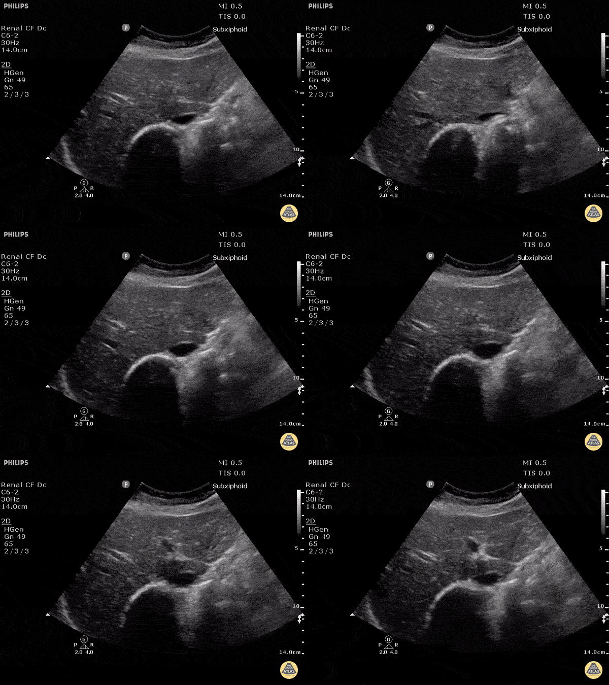
Inferior Vena Cava in Hypovolemia (Transverse)
Justin Bowra MBBS, FACEM, CCPU Emergency Physician, RNSH et al.
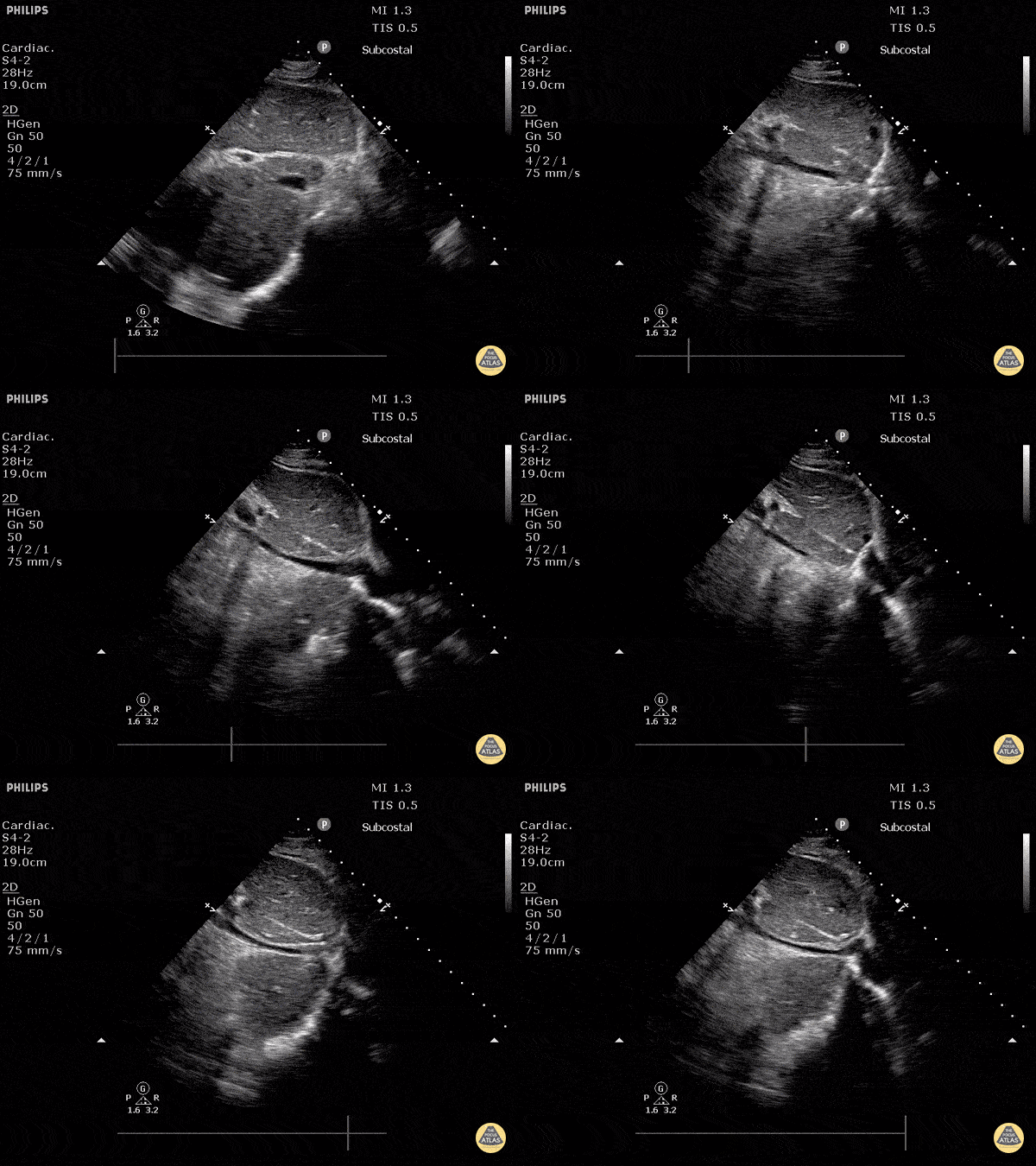
Inferior Vena Cava in Hypovolemia
Justin Bowra MBBS, FACEM, CCPU Emergency Physician, RNSH et al.
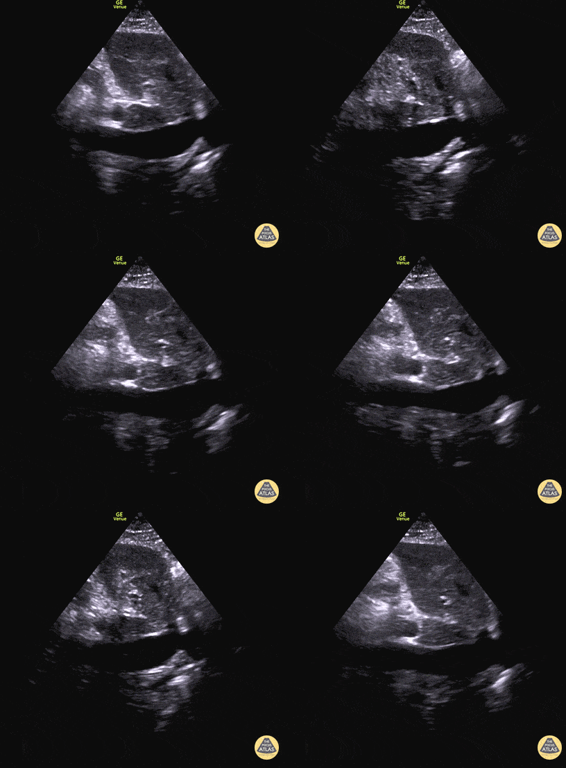
Plethoric IVC in Biventricular Failure
50 y/o M PMH methamphetamine abuse presents in respiratory failure with cool, mottled skin and poor capillary refill. Patient was tachycardic, hypothermic with multiple signs of end organ dysfunction. POCUS quickly narrowed the differential by demonstrating clear, severe, reduced ejection fraction by visual estimation and a plethoric IVC without respiratory variation. Patient was ultimately started on dobutamine and transferred to the MICU for treatment of cardiogenic shock.
Dr. Jaclyn Walker, Dr. Matthew Riscinti - Denver Health Emergency Medicine
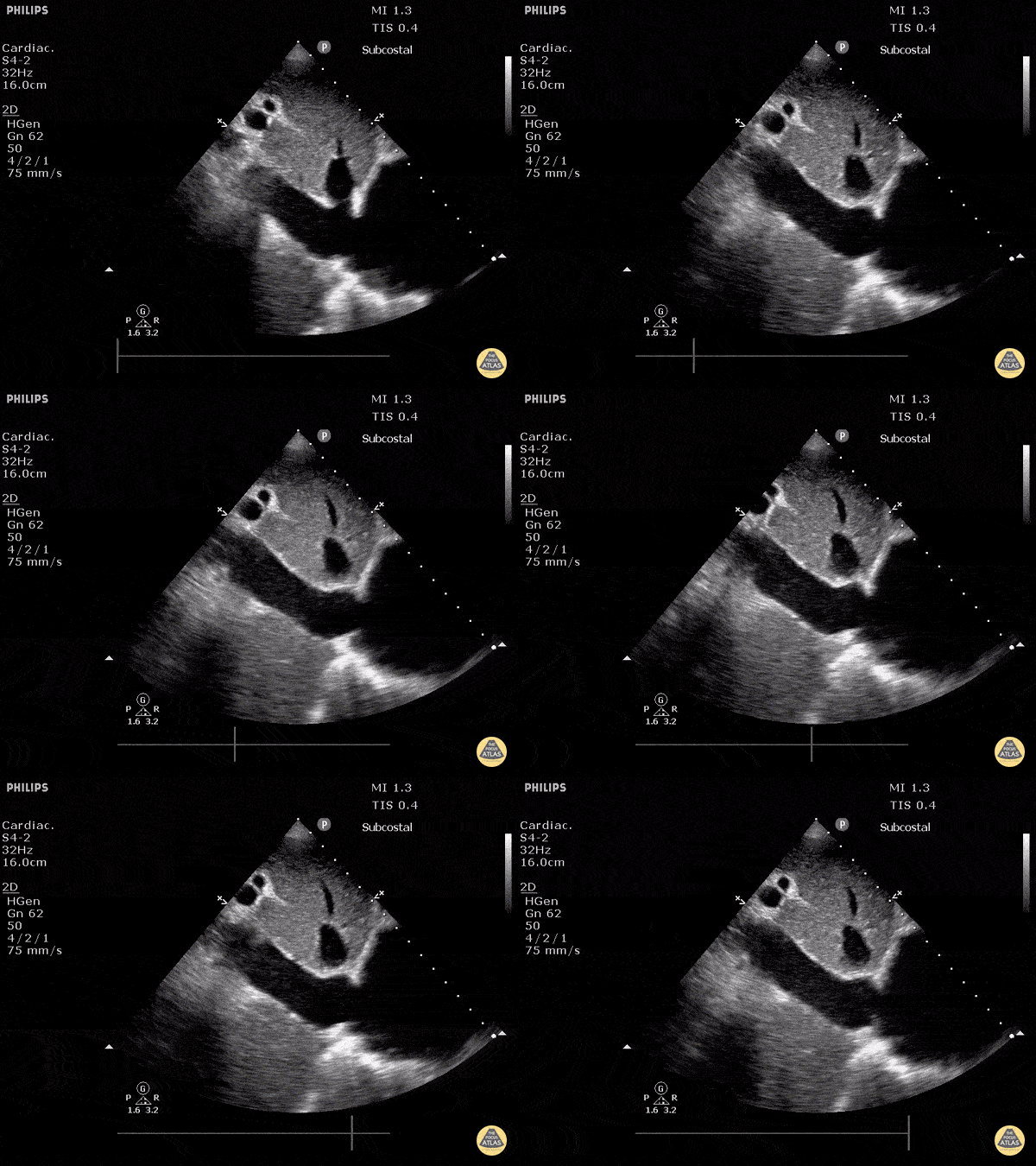
Plethoric Inferior Vena Cava
Justin Bowra MBBS, FACEM, CCPU Emergency Physician, RNSH et al.
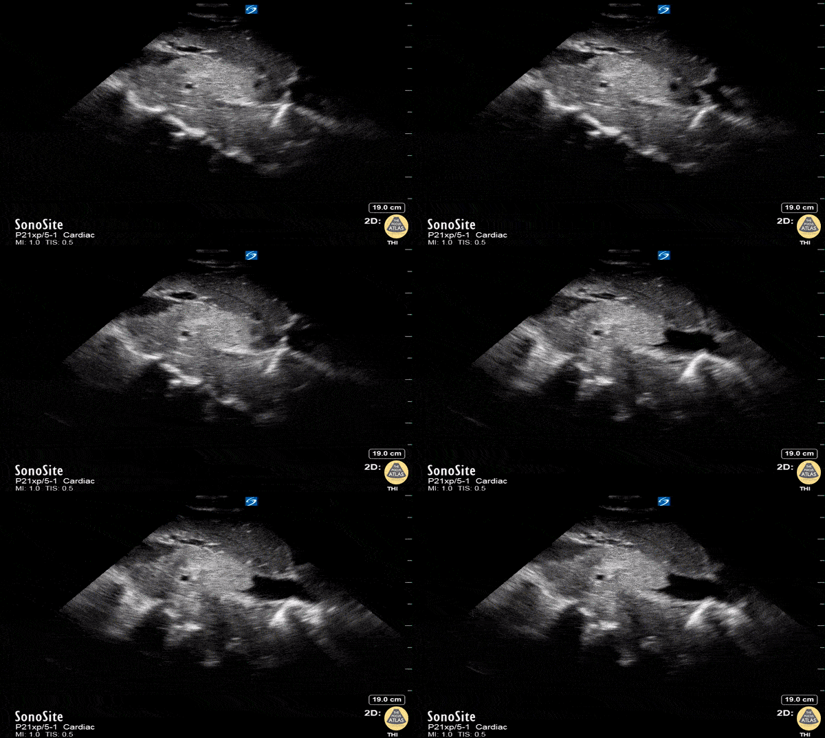
IVC Thrombus
A 30-year-old man presented with shortness of breath. Normally fit and well with no past medical history apart from a lump in right axilla. Observations were stable apart from a high respiratory rate. Physical examination revealed more lymph nodes in the groin. Working diagnosis of Lymphoma and sent for a CT chest, abdomen and pelvis. In the interim, point of care ultrasound showed clot in the IVC. Confirmed on CT scan. Patient was ultimately diagnosed with metastatic testicular cancer and tumour thrombus in the IVC, which was managed conservatively with anticoagulation.
Dr Parmy Deol, Emergency Physician, Chelsea and Westminster hospital, London

Significantly Enlarged IVC in Acute Heart Failure
This is a subxiphoid long axis view of the IVC demonstrating significant enlargement with minimal collapse with respiratory variation. The hepatic vein is also seen entering the IVC and is noted to be quite distended consistent with very high filling pressures. Justin Bowra MBBS, FACEM, CCPU Emergency Physician, RNSH et al. (Dr. Vahtrick)


