Occult pneumothorax: tube or observe?
Tareq Azad, MD
This post is a collaboration between on The POCUS Atlas and Clinical Monster. If you're interested in collaborating on a POCUS oriented topic, contact us.
It’s another day at your local hospital. You’re nearing the end of your shift and wrapping up your cases. Your mind is already starting to wander with thoughts of what you’ll do on vacation. Suddenly, you hear some commotion stir through the ED -- TRAUMA CODE to the hospital LOBBY.
A few minutes later, you’re in the resuscitation bay with the patient. He is a 26-year-old male with a stab wound to the left lateral chest, inferior to the nipple. As IVs and O2 are being started, and the patient is being put on the monitor, you quickly run through your ABCs. The patient’s airway, breathing, and circulation are intact with clear breath sounds bilaterally. Secondary survey shows normal vital signs and a single 1 cm puncture wound to the L chest at the anterior axillary line. The patient doesn’t remember what the weapon looked like or any other details regarding the injury. You obtain a STAT portable chest radiograph, which appears normal.
Despite this normal appearing chest radiography, you still have a high suspicion for some underlying pulmonary injury. Being the clinical monster that you are, you quickly grab the ultrasound machine for some point-of-care-ultrasonography (POCUS).
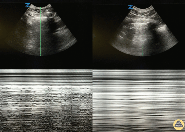
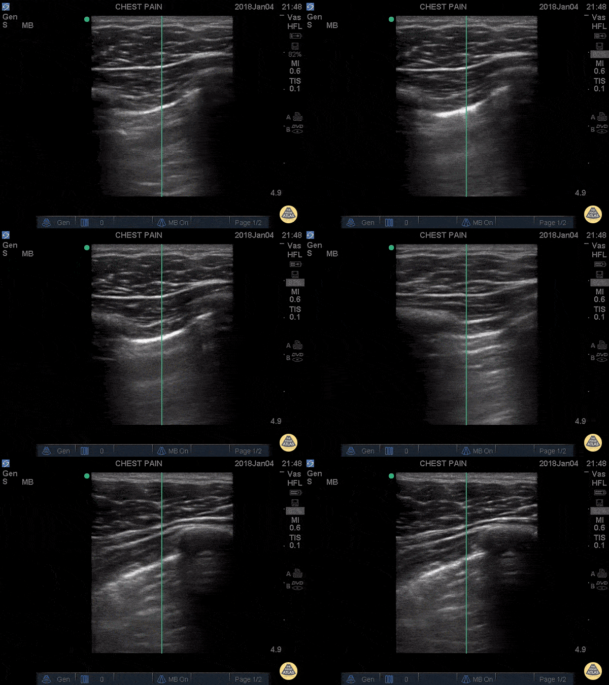
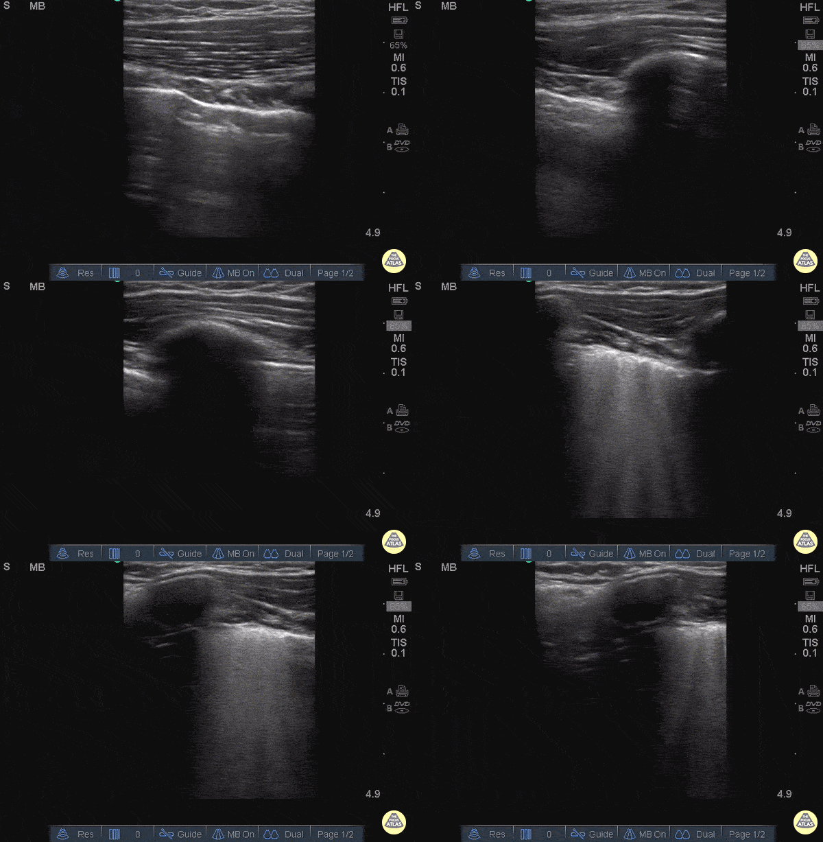
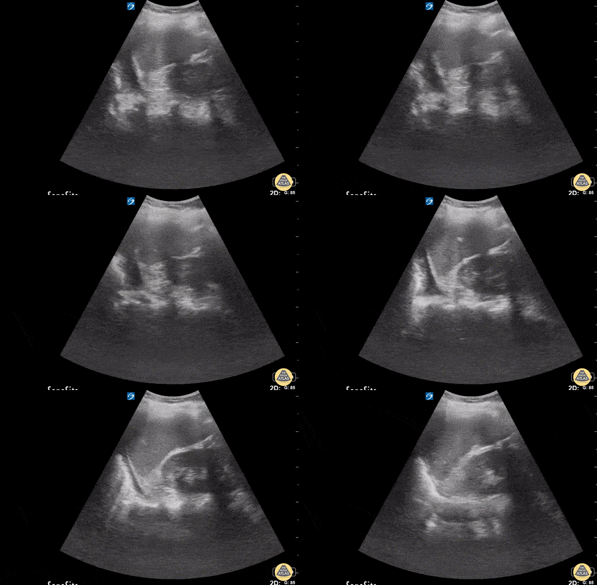
As you perform your eFAST, you find an area without lung sliding or B-lines that is transitioning into view of normal lung sliding. This is known as a lung point, a highly specific (nearing 100%) sonographic finding for pneumothorax. This point represents the physical boundary of the pneumothorax, outlining the area where the parietal and visceral pleura separate. While the absence of lung sliding itself is a fairly sensitive indicator of pneumothorax, pleural adhesions (such as in malignancy) or pleural blebs (such as seen in COPD) may also lead to decreased lung sliding, thereby decreasing specificity.
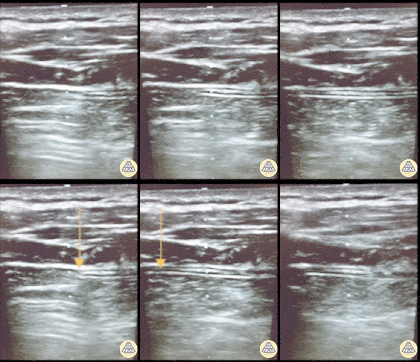
Another example of lung point from the POCUS Atlas Trauma section. Lung point indicates the transition point between normal pleura with normal lung sliding (on the left side of the image) and where there is air disrupting the pleural space with decreased lung sliding (on the right side of the image). Lung point is a highly specific finding indicating a pneumothorax. Dr. Sathya Subramaniam, Pediatric EM Fellow - Kings County/SUNY Downstate
With your lung point visualized and post-test probability high, you order a confirmatory chest CT and find a 20% pneumothorax. This is an occult pneumothorax - a pneumothorax that is not initially seen on supine chest radiograph but later found on advanced imaging such as CT.2 Now what?
Acute management of occult traumatic pneumothorax is an area of controversy. The two management options are 1) observation or 2) immediate thoracostomy. Tube thoracostomy, while often life-saving, is sometimes a morbid procedure with complications including improper tube placement, injury to surrounding structures or vessels, and infection in up to 20% of patients.3,4 Management of an unstable patient is often straightforward with immediate surgical intervention, but the well-appearing patient often brings the question of whether thoracostomy is truly warranted.
A systematic review by Yadav et al. looked at ED trauma patients of all ages with occult pneumothorax at initial presentation after blunt or penetrating trauma. . The study searched MEDLINE, EMBASE, and COCHRANE Library databases and found 3 RCTs with 101 adult patients in total after inclusion criteria were applied. Occult pneumothorax was defined as a pneumothorax not seen on initial supine radiography but later found on abdominal or chest CT. The intervention group were those patients that were observed at presentation, the comparison group were those patients that had tube thoracostomy or any other form of indwelling pleural drainage. . This study looked at outcomes of mortality, progression of pneumothorax, development of pneumonia or empyema, and length of stay in hospital. This study showed that observation of patients with occult pneumothorax results in similar outcomes compared to those treated with tube thoracostomy. However, a particular point of contention is whether those patients placed on positive pressure ventilation are at higher risk for progression of occult pneumothorax to tension pneumothorax, and thus should be more aggressively managed at outset. One limitation of the study was that while all three trials reported to be randomized, only one trial included data regarding their randomization method. None of the three trials determined their sample size based on a priori based clinical outcomes, thus outcomes of the individual studies may have been due to lack of power and nuanced differences between the groups may have been missed. None of the three selected RCTs had sufficient statistical power to be able to form statistically significant conclusions.
In conclusion, be cognizant of lung points - don’t be fooled if you find normal lung sliding surrounding a point of what appears to be absent lung sliding. In the management of occult traumatic pneumothorax - when the patient is stable and otherwise well-appearing - some studies show that observation is a reasonable approach. If you see a pneumothorax on POCUS, it does not necessarily mean you need to immediately place a chest tube, but it does help you to immediately get prepared for one.
Tareq Azad, MD
Peer Reviewers: Kyle Kelson, MD and Matt Riscinti, MD
Faculty Reviewer: Ian deSouza, MD and John Kilpatrick, MD
This post is co-hosted on The POCUS Atlas and Clinical Monster. The post on Clinical Monster can be found here.
More Pneumothorax POCUS cases from our Trauma Atlas
References:
Hew M, Tay TR. The efficacy of bedside chest ultrasound: from accuracy to outcomes. European Respiratory Review 2016;25(141):230–46.
Mowery NT, Gunter OL, Collier BR, et al. Practice Management Guidelines for Management of Hemothorax and Occult Pneumothorax. The Journal of Trauma: Injury, Infection, and Critical Care 2011;70(2):510–8.
Rivera L, O’Reilly EB, Sise MJ, et al. Small Catheter Tube Thoracostomy: Effective in Managing Chest Trauma in Stable Patients. The Journal of Trauma: Injury, Infection, and Critical Care 2009;66(2):393–9.
Etoch SW. Tube Thoracostomy. Archives of Surgery 1995;130(5):521.
Yadav K, Jalili M, Zehtabchi S. Management of traumatic occult pneumothorax. Resuscitation 2010;81(9):1063–8.


