
Other Cardiac Pathology

Migrated Venous Stent Traversing Tricuspid Valve
Middle aged male with recent right iliac DVT s/p thrombectomy and venous stent placement is found to have asymptomatic migration of stent into the right heart. In the subxiphoid view, the hyperechoic coiled-appearing stent is seen traversing the tricuspid valve. Percutaneous attempt at removal was unsuccessful, so he ultimately underwent open-heart surgery.
Contributed by: Eric Reid, MD

Clot Within Right Atrium
This patient initially presented post-operatively to the emergency department with complaints of dyspnea. From this subxiphoid view of the heart, we can see a hyperechoic structure suggestive of clot formation. An alternative view of this clot viewed from the right atrium and IVC junction can be seen here, termed “clot-in-transit”. The patient was subsequently diagnosed with a DVT that extended into their central femoral vein.
Image courtesy of Robert Jones DO, FACEP @RJonesSonoEM
Director, Emergency Ultrasound; MetroHealth Medical Center; Professor, Case Western Reserve Medical School, Cleveland, OH
View his original post here

POCUS during Resuscitation
This is a PLAX view obtained during CPR . It reveals adequate compressions with the LV “squashing” and obliterating the LV lumen; then subsequently the LV lumen reappearing and recoiling, which allows the LV to refill.
Renato Tambelli, @JediPocus

LV thrombus, parasternal short axis
Parasternal short axis view of a 47yo with PMH non-obstructive CAD incidentally found to have mildly reduced LV systolic function and an echogenic mass in LV extending to outflow tract. Determined to be a large LV thrombus of unknown etiology.
Andrew Balster, MD
Paul Musgrave, MD (OHSU IM POCUS fellow)
@POCUSaurusDx

LV mass/thrombus
Parasternal long axis view of a 47yo with PMH non-obstructive CAD incidentally found to have mildly reduced LV systolic function and an echogenic mass in LV extending to outflow tract. Determined to be a large LV thrombus of unknown etiology.
Andrew Balstera, MD
Paul Musgrave, MD (OHSU IM POCUS Fellow)
@POCUSaurusDx

Pacer Wire Perforation
Patient with chest pain with recent history of pacemaker placement. Subxiphoid view reveals the pacer wire in the right ventricle piercing the wall, causing a pericardial effusion.
Image courtesy of Robert Jones DO, FACEP @RJonesSonoEM
Director, Emergency Ultrasound; MetroHealth Medical Center; Professor, Case Western Reserve Medical School, Cleveland, OH
View his original post here

Venous Air Embolism
Pictured here is an apical four chamber view of the heart in a patient who presented for evaluation of dyspnea and was found to be in atrial fibrillation with RVR. POCUS is most notable for the presence of gas bubbles throughout the right atrium and right ventricle. Symptoms of venous air embolism can range from asymptomatic to cardiovascular collapse and death. The presence of venous air embolism may occur due to head or neck surgery, chest trauma, hemodialysis, central vein cannulation, high pressure mechanical ventilation, or thoracentesis.
Rupinder Sekhon, MD & Peter Biggane, MD
Central Michigan University, Emergency Medicine

Bi-Ventricular Thrombi
A 30-year-old male with history of idiopathic heart failure was hospitalized and on mechanical ventilation for COVID-19. POCUS included this subcostal image notable for severe bi-ventricular dysfunction complicated by thrombus formation in the left and right ventricles.
Patricia Lopes, Resident of Emergency Medicine at Fortaleza-CE, Brazil
@patylopes90
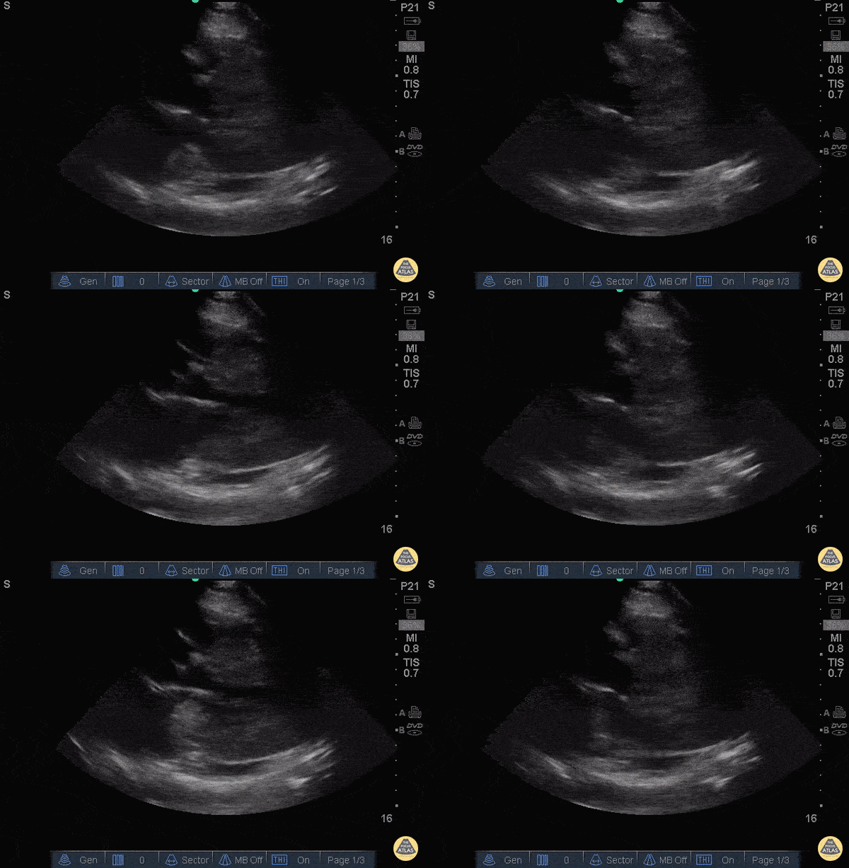
A Fib with Left Atrial Thrombus (parasternal long)
40 y/o M with atrial fibrillation, off his anticoagulation, presented with shortness of breath and was found to have a left atrial thrombus on POCUS in this parasternal long view.
Differentiating a thrombus from an atrial myxoma is difficult, but seeing the object tumble and shoot around the atrium, in the setting of afib, is both dramatic and suggestive of a thrombus. Unfortunately, timing does not differentiate the two since the average myxoma can grow at up to 0.5cm/month.
The patient was admitted to the cardiac intensive care unit with full resolution of the thrombus after anticoagulation.
Dr. Bryan Jarrett and Dr. John Kilpatrick - SUNY Downstate/Kings County Emergency Medicine
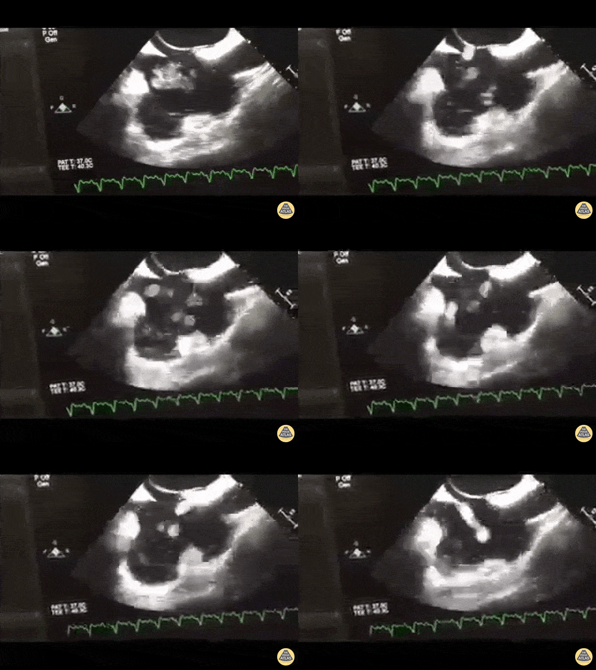
Thrombus in Transit
Patient in shock. TEE performed as TTE views not obtainable. Midesophageal bicaval view demonstrating what we've dubbed the "RA washing machine".
WCUME 2017 Submission for "Best POCUS" Dr. Tom Jelic (@TomJelic) Winnipeg, Canada
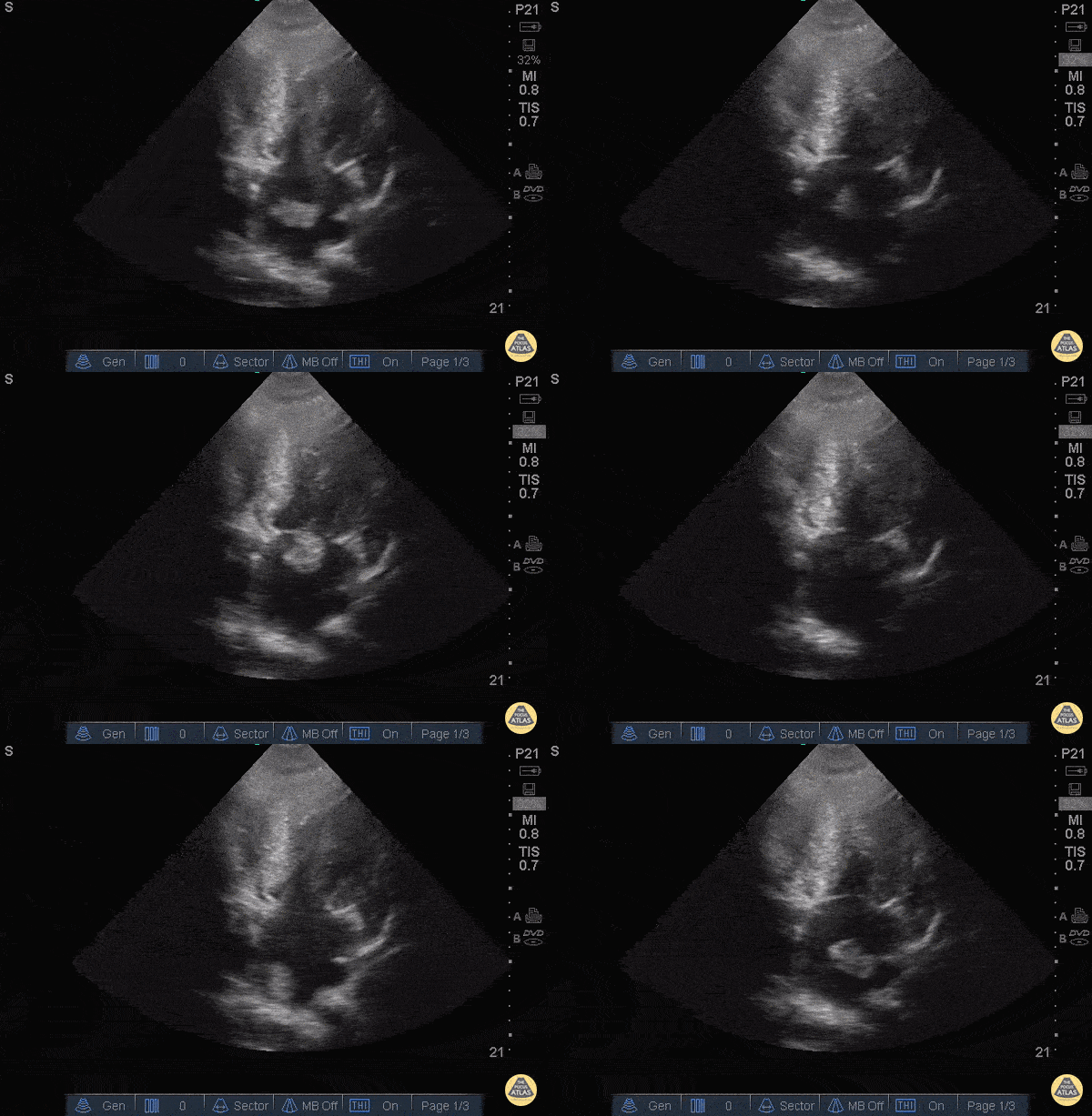
A Fib with Left Atrial Thrombus
40 y/o M with atrial fibrillation, off his anticoagulation, presented with shortness of breath and was found to have a left atrial thrombus on POCUS in this apical view.
Differentiating a thrombus from an atrial myxoma is difficult, but seeing the object tumble and shoot around the atrium, in the setting of afib, is both dramatic and suggestive of a thrombus. Unfortunately, timing does not differentiate the two since the average myxoma can grow at up to 0.5cm/month.
The patient was admitted to the cardiac intensive care unit with full resolution of the thrombus after anticoagulation.
Dr. Bryan Jarrett and Dr. John Kilpatrick - SUNY Downstate/Kings County Emergency Medicine
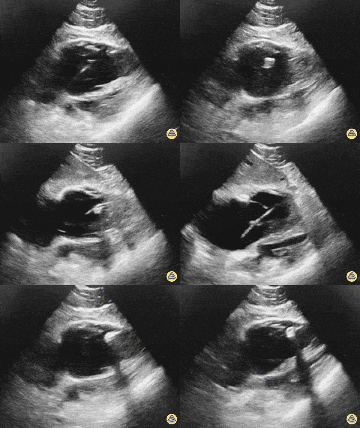
Transvenous Pacemaker Guidance
WCUME 2017 Submission for "Novel Indication"
Confirming Transvenous Pacer Placement with POCUS.
Dr. Sarah Medeiros, MD - Sacramento, CA
The Saline Flush/Bubble Test
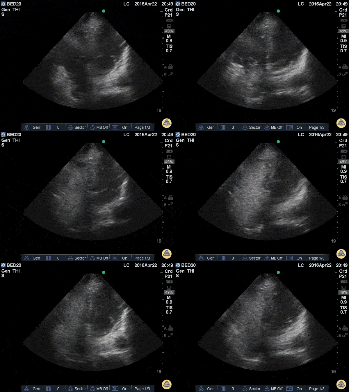
The included clip demonstrates an apical 4 chamber view with saline bubbles in the right atrium and right ventricle after a quick 10ml flush through a right IJ central line.
56 y/o M with pneumonia, respiratory distress, intubated, and hemodynamically unstable was started or levophed through a peripheral IV while a right IN was started. The portable x-ray tech wa missing but rapidly flushing saline through the IJ during apical 4 clearly demonstrates saline the R circulation
After placing central lines, confirmation by chest x-ray is not always available. When the patient is unstable, delayed use can be consequential. Visualizing the saline swirling in the right side of the heart immediately after a flush is highly predictive of proper catheter tip placement.
Weekes AJ et al. Central vascular catheter placement evaluation using saline flush and bedside echocardiography. Acad Emerg Med. 2014 Jan;21(1):65-72. doi: 10.1111/acem.12283.
Leon Chen, NP – Critical Care Medicine Service; Department of Anesthesiology and Critical Care Medicine; Memorial Sloan Kettering Cancer Center; New York, NY
AV Canal Defect
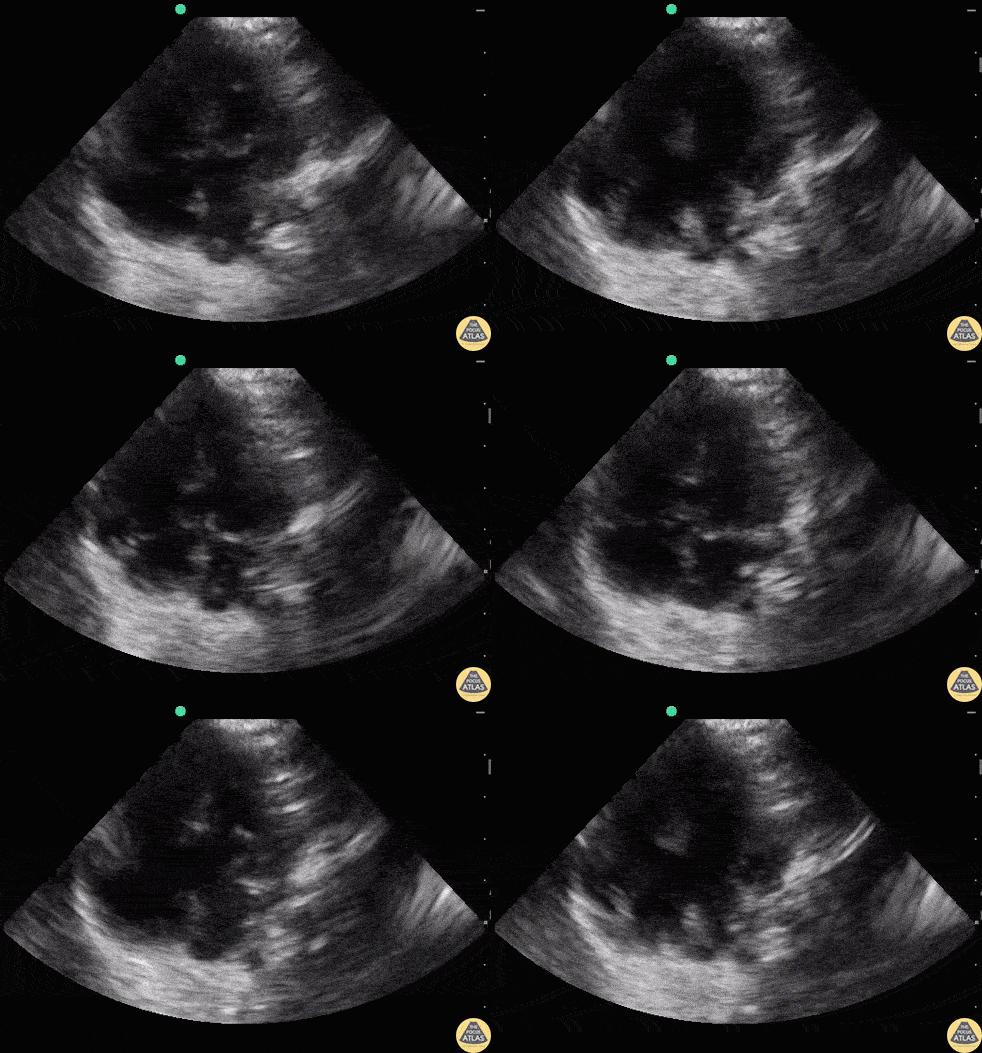
3 month old recent immigrant to the United States with Trisomy 21, presenting with nasal congestion, cough, respiratory distress, with mild abdominal breathing, presumably with bronchiolitis. POCUS was performed to assess the lungs and heart.
POCUS revealed a complete AV canal defect which is often seen in Trisomy 21. The interventricular septum has a free end with a common AV valve present and a free end of the ostium primum with ASD.
Dr. Sathya Subramaniam - Kings County/SUNY Downstate - Pediatric EM Fellow
Truncus Arteriosus
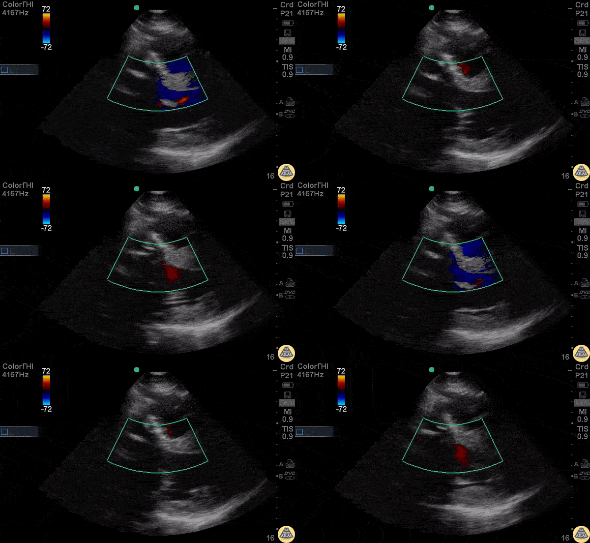
Parasternal long view with color doppler of a young male presenting with chest pain and a history of surgically corrected truncus arteriosus including an aortic valve replacement. Notable here is the regurgitant jet just above the aortic outflow tract. The etiology of this jet is uncertain but could represent a persistent VSD despite correction.
Dr. Bryan Jarrett - Kings County Emergency Medicine
Truncus Arteriosus
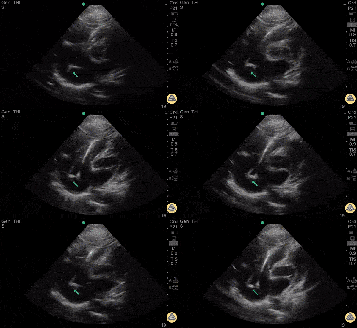
35 y/o M PMH truncus arteriosus s/p multiple surgeries, bioprosthetic aortic valve, AICD, and recent treatment for endocarditis presents with rectal bleeding and syncope in setting of warfarin use for his chronic DVTs. INR was found to be 1.0. POCUS with pacemaker leads with possible clot vs vegetation attached to the tip. Concern for thrombus given pt's normal INR vs possible infected pacemaker vegetation.
Dr. Bryan Jarrett - Kings County Emergency Medicine
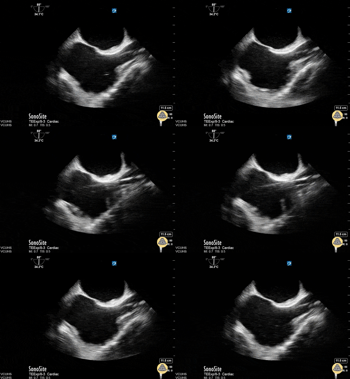
TEE - ECMO Placement Confirmation
TEE confirmation of ECMO Cannula done at VCU.
Dr. Stenberg

Free Peritoneal Fluid seen with Falciform Ligament
50s F found to be in cardiac arrest by EMS in the setting of 3 days hematemesis, achieved ROSC, and this image was seen on POCUS performed by EMS while packaging for transport. This subxiphoid view demonstrates the presence of organized cardiac activity with no large pericardial effusion, but free peritoneal fluid is seen adjacent to the liver, superficial to the diaphragm in this image, and the falciform ligament is also briefly seen.
Zachary Hutchins
South Metro Fire Rescue, Centennial, CO

Cardiac Amyloidosis
Parasternal long axis taken in a patient with clinical suspicion of cardiac amyloidosis.
Maxime Gautier
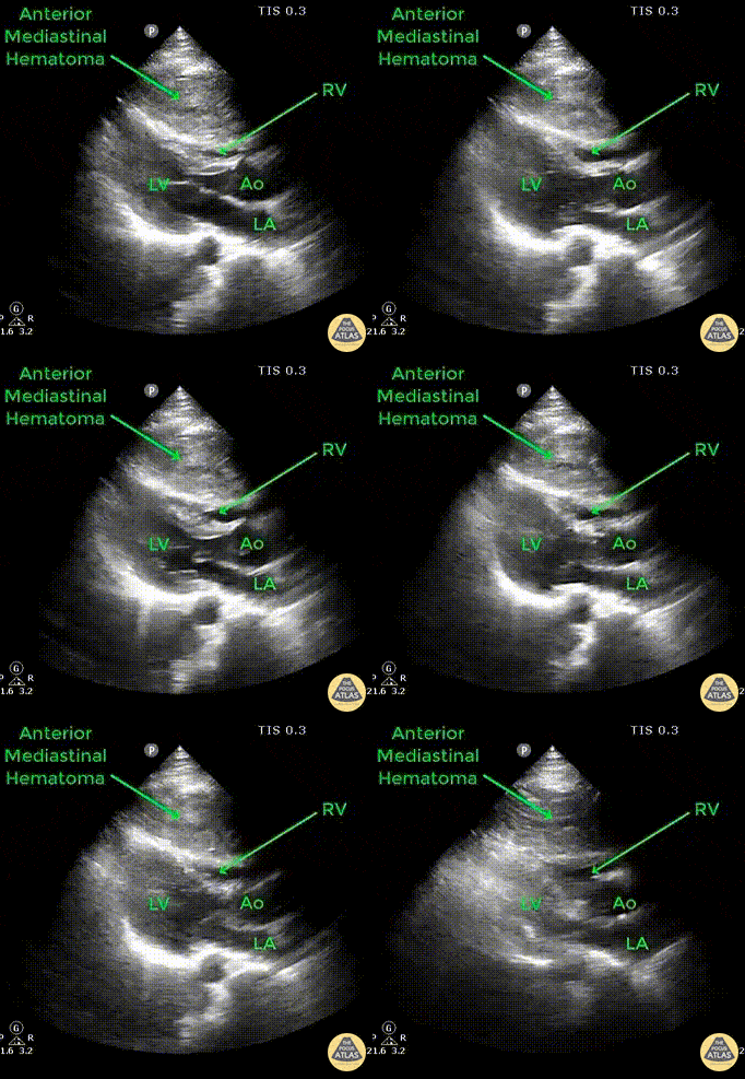
Anterior Mediastinal Hematoma
Pt presents to ED following an unrestrained head-on MVC with blunt force trauma to chest. Compressing the RV is an anterior mediastinal hematoma seen in this parasternal long axis view secondary to a sternal fracture.
Image courtesy of Robert Jones DO, FACEP @RJonesSonoEM
Director, Emergency Ultrasound; MetroHealth Medical Center; Professor, Case Western Reserve Medical School, Cleveland, OH
View his original post here

Retained Intracardiac Air
A 70 year old male presents 3 days post radiofrequency ablation for atrial fibrillation and atrial flutter. Presented to emergency department with evidence of septic shock, encephalopathy, and hypoxic respiratory failure. The above image shows evidence of air bubbles within the right atrium and ventricle, thought to be iatrogenic in nature after his recent cardiac procedure possibly due to necrotizing infection of the myocardium versus atriotracheal fistula or atrioesophageal fistula. The patient was placed in reverse Trendelenburg position (to mitigate potential embolus to cerebral venous circulation). Fortunately, following flight transfer to another facility, the intracardiac air was no longer visible on CT or echocardiography.
Ian Keck
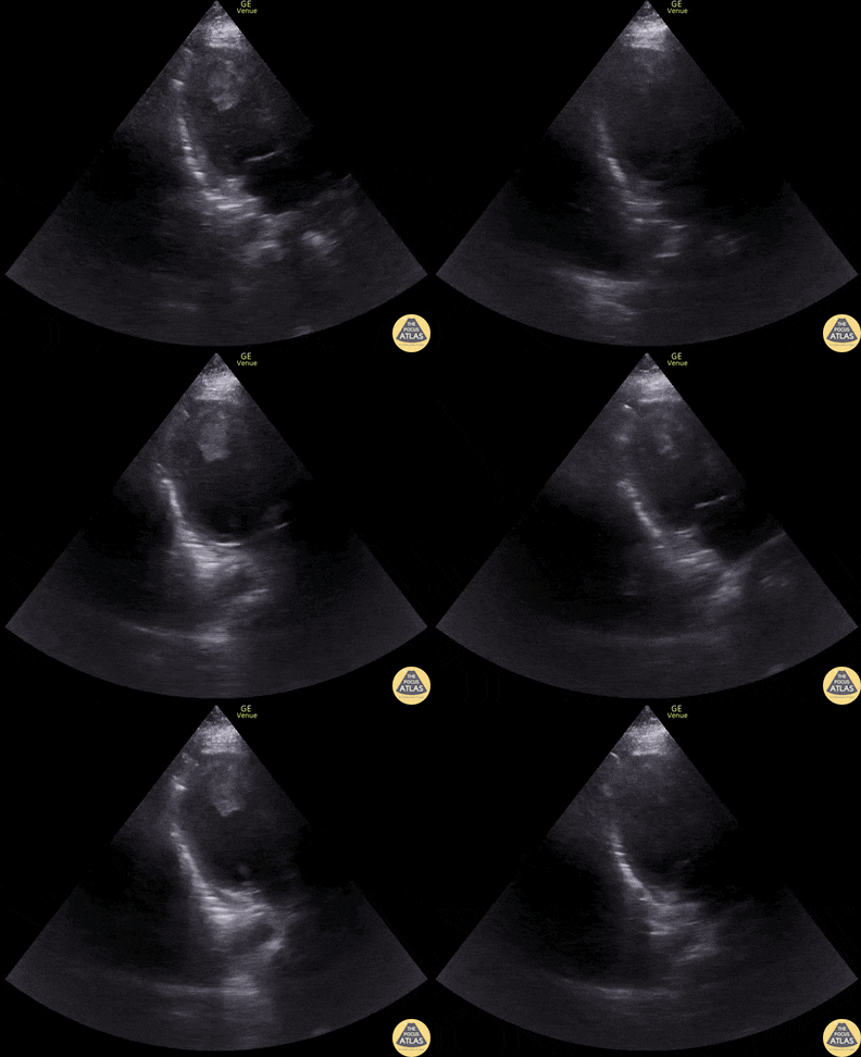
LV Thrombus
40s M with PMH HTN/HLD with no known CHF history presented with BLE edema, dyspnea, orthopnea, and weight gain. POCUS performed shortly after arrival showed markedly reduced LVEF with apical LV thrombus. The patient was initiated on a heparin drip and admitted, where formal TTE and right and left heart caths confirmed HFrEF due to ischemic cardiomyopathy and 3v CAD. Ultimately this patient improved with medical therapy but had an acute massive embolism event to b/l femoral arteries, b/l renal arteries, and SMA about 1 week into his hospitalization despite being on heparin drip while bridging to warfarin. He went to the operating room with vascular surgery and interventional cardiology for extensive thrombectomies as well as angiography but ultimately died after requiring resection of ischemic bowel and later suffering a large hemispheric embolic CVA.
Dr. Kathleen Joseph, PGY-4, Denver Health Residency in Emergency Medicine
Dr. Cailin Frank, Fellow, Denver Health Ultrasound Fellowship

Hypertrophic Cardiomyopathy (Long)
Justin Bowra MBBS, FACEM, CCPU Emergency Physician, RNSH et al. (Dr. Kaveh Kaynama)

Bubble Study for CVL Verification
A 33-year-old female presented with cocaine-induced chest pain, dyspnea, and clinical evidence of cariogenic shock. CVL was emergently placed and location confirmed via POCUS Bubble Study. Ultrasound verification of CVL placement is possible by visualizing microbubble artifact in the right atrium following injection of saline through the distal port of the CVL.
Dr. Victor Bang. Emergency Physician at Hospital das Clínicas de Marília. Co-founder of Pocus Jedi.
@vmjbang


